
Transplant Pathology
Contributed by Hugo Kaneku, MD and Marta Minervini, MD
Department of Patholgy, Division of Transplantation Pathology, University of Pittsburgh Medical Center
Clinical History
The patient is a young female who presented at her local hospital with 2 weeks of fatigue, fever, abdominal pain, nausea, vomiting and jaundice. Blood workup at that time was positive for IgM anti-HAV and a diagnosis of acute hepatitis A was established. She presents 3 months later with persistent jaundice, nausea, vomiting and elevated liver enzymes (AST 956, ALT 455, alkaline phosphatase 88, total bilirubin 16.5). Other relevant labs showed: INR 2.4, positive anti-smooth muscle antibodies (ASMA), elevated IgG level of 2530 and negative serologies for HBV and HCV, anti-nuclear (ANA) and anti-mitochondrial-M2 antibodies (AMA-M2). A liver biopsy is performed.
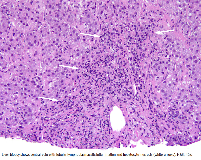
Biopsy Findings
The biopsy shows portal, periportal and lobular hepatitis with significant inflammation and hepatocyte loss. In the patient's clinical context, the differential diagnosis included acute HAV infection and autoimmune hepatitis (AIH). The distinction between these two etiologies is problematic given that there are many overlapping histological findings including marked plasma cell rich interface activity, portal and lobular inflammation, and hepatocellular necrosis. Also, serological studies support both entities. Further complicating the situation is the observation that HAV hepatitis can trigger the onset of AIH in susceptible individuals (1). There is also periportal and pericellular fibrosis with the latter concerning for a previous episode of steatohepatitis and/or chronic autoimmune hepatitis, upon which the acute necroinflammatory process is superimposed.
This case was discussed at the weekly Interdepartmental conference with the hepatology team and prompted the clinical discussion on the potential etiology with the final agreement that this case represented an HAV infection with an unusual presentation. Therapeutically, the use of steroids was discussed as this would be applicable for either diagnosis. The possibility of using HAV immunoglobulin was also discussed.
During the hospitalization, the patient developed encephalopathy and liver failure, requiring a liver transplant. She received a living-unrelated liver from an altruistic donor. Microphotographs of relevant areas from the native liver are shown below.
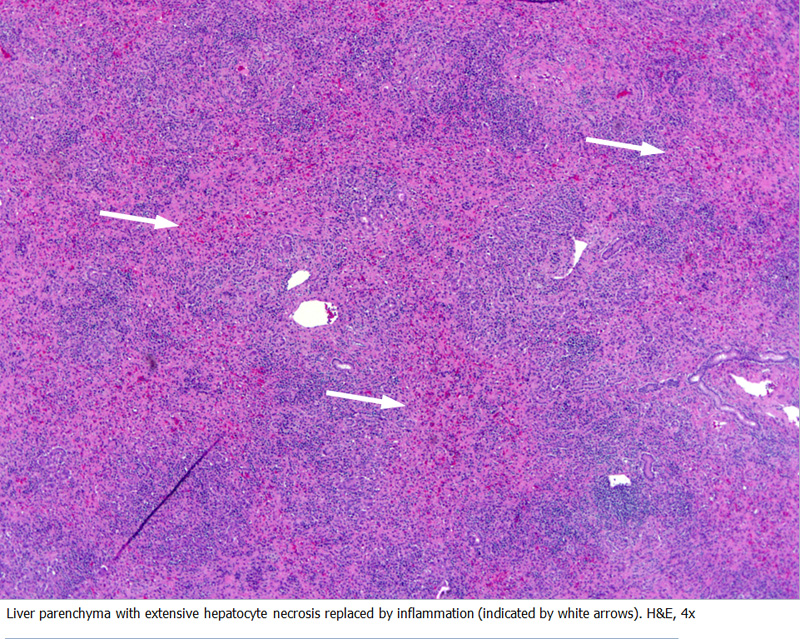
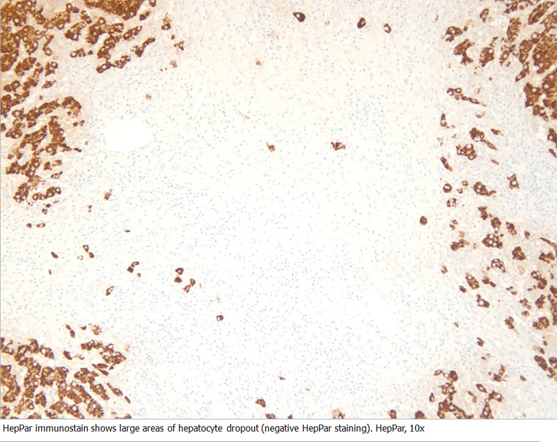
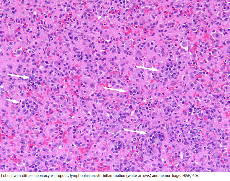
Examination of the native liver explant shows panlobular necrosis involving about 60% of the liver, associated with hemorrhage, lymphoplasmacytic inflammation and mild ductular reaction. Areas of more preserved liver showed moderate interface activity with many plasma cells, perivenulitis, moderate hepatocanalicular cholestasis and focal confluent necrosis. Again, because of overlapping features, the distinction between HAV and AIH is challenging.
About a week after transplant, the LFTs were still elevated and a liver allograft biopsy was performed.
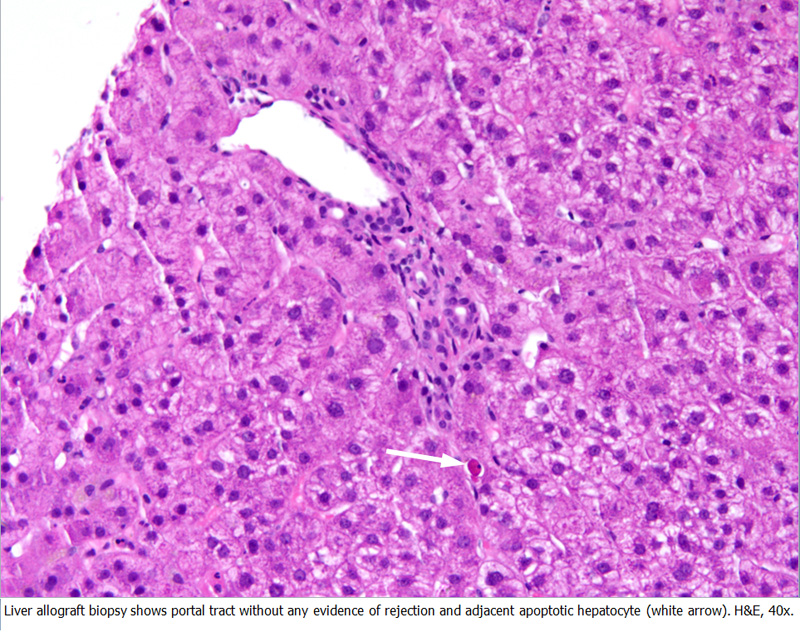
The liver biopsy shows no signs of T-cell mediated or antibody mediated rejection. There were occasional apoptotic hepatocytes with mild hepatocanalicular cholestasis and no fibrosis. These findings were interpreted as likely representing suboptimal vascular inflow, although similar findings had been reported in the few cases published in the literature of recurrence of hepatitis A virus infection after liver transplantation (2-4). Some common findings in these reported cases included portal and lobular inflammation, apoptotic hepatocytes and cholestasis. These cases were positive for HAV RNA and anti-HAV IgM, and seroconversion to anti-HAV IgG was seen after adjusting the immunosuppressive medications.
Subsequently, the patient underwent splenic artery embolization expecting that some of the splenic blood flow will be redirected to the liver and alleviates the blood inflow issue. The patient was discharged home 2 weeks after her liver transplant and is doing well.
Discussion
This is an unusual presentation of acute HAV infection causing liver failure and requiring a liver transplant. HAV usually presents as an acute infection ranging from mild to severe disease, does not have a chronic clinical form, and less than 1% can develop liver failure. About a third of patients with acute liver failure secondary to HAV need a transplant, while the other two thirds recover spontaneously (5). Preexisting liver conditions like non-alcoholic and alcoholic liver disease makes individuals more susceptible to develop liver failure. This is consistent with what we observed in our patient, as the possibility of an underlying chronic steatohepatitis (as mentioned in the first biopsy) could not be entirely excluded. Further complicating the situation is the empiric observation that HAV hepatitis can trigger the onset of AIH particularly in susceptible individuals. Finally, recurrent HAV infection after liver transplantation is even rarer and only a handful of cases have been reported in the literature.
References
- Vento S, et al. Identification of hepatitis A virus as a trigger for autoimmune chronic hepatitis type 1 in susceptible individuals. Lancet 1991, vol 337, issue 8751, 1183-1187
- Fagan E, et al. Persistence of hepatitis A virus in fulminant hepatitis and after liver transplantation. Journal of Medical Virology 30: 131-136 (1990).
- Eisenbach C, et al. Recurrence of clinically significant hepatitis A following liver transplantation for fulminant hepatitis A. Journal of Clinical Virology 35 (2006) 109-112.
- Park GC, et al. Intractable recurrent hepatitis A virus infection requiring repeated liver transplantation: a case report. Transplantation Proceedings, 42, 4658-4660 (2010).
- Taylor RM, et al. Fulminant hepatitis A virus infection in the United States: incidence, prognosis, and outcomes. Hepatology 2006; 44: 1589-1597.