
Soft Tissue Pathology
Contributed by Virginia Miller, MD and Jon M. Davison, MD, Department of Pathology, University of Pittsburgh Medical Center
Clinical History
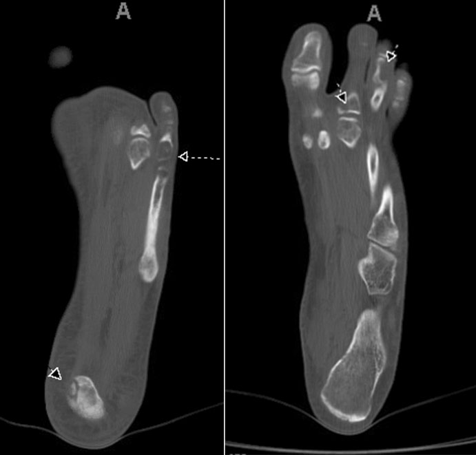
A Caucasian male in his early 20's presented with a cutaneous, soft tissue mass of the right lower extremity as well as vascular abnormalities of the mid-3rd phalange. Subsequent pathologic fracture of the proximal 2nd phalange, led to the discovery of multiple osseous lesions of the left foot (below). Excision of the lesions showed similar histologic features in both the soft tissue and bone sites. Long-term follow up was characterized by multiple bone (tibia) and soft tissue recurrences within the tissues of the right lower limb.
Imaging
Histology
Low-power cutaneous lesion
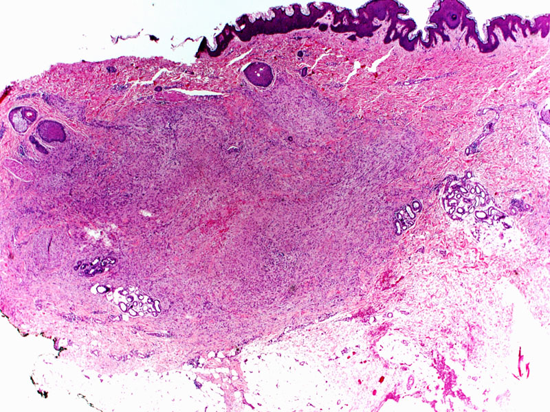
Periphery of cutaneous lesion with infiltrative border
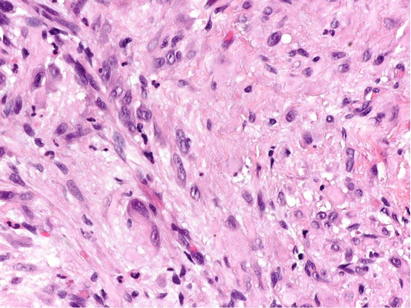
High-power cutaneous lesion with cytologic/nuclear detail
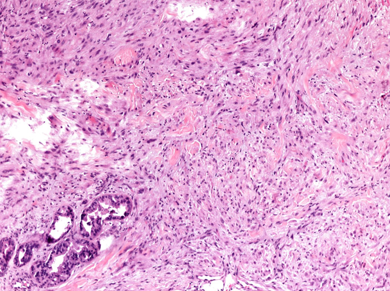
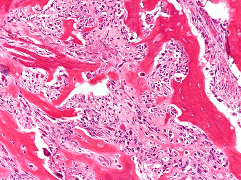
High-power bony lesion, post decal (2nd phalange)
Stains
Molecular Findings
Fish
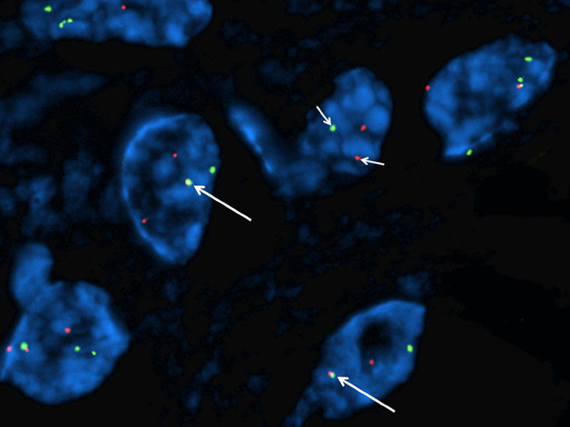
FOSB-1/SERPINE-1 t(7;19) gene fusion.
Final Diagnosis
Pseudomyogenic Hemangioendothelioma (PHE)
Discussion
Pseudomyogenic hemangioendothelioma (PHE) is a vascular lesion of intermediate grade with a propensity to affect younger males (2nd -5th decade, M:F 5:1) often as multi-focal disease with cutaneous, soft tissue and/or bone involvement. Commonly noted in the limbs, especially lower extremity, the nodules may be painful or asymptomatic and manifest within multiple tissue planes in a regional fashion. Radiologic workup with PET should be pursued to fully detect deeper lesions and evaluate extent of disease as a significant percent of those presenting with superficial lesions have concurrent bone involvement. Radiographically, lesions are multiple, discrete, and lucent.
Grossly, the nodules are small (1-2 cm) and solid, appearing well-circumscribed. Histologically, they present as sheets of plump/epithelioid to spindled cells with abundant, eosinophilic cytoplasm, large nuclei with open, vesicular chromatin. Microscopic margins are ill-defined with tendrils of tumor weaving throughout surrounding collagen bundles. Neither nuclear pleomorphism nor mitoses are prominent. Infiltrating neutrophils may be conspicuous in the background and small foci of necrosis may be observed.
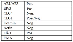
The intermediate grade of PHE reflects a behavior of rapid loco-regional recurrence, but distant metastases are exceptionally rare. Conservative resection is the preferred therapy with chemotherapeutic options as adjunct. Diagnosis is definitive with immuno-molecular work up, however PHE is keratin (AE1/AE3) positive, which may prove a diagnostic pitfall considering its vascular origin and the histologic differentials. ERG displays strong nuclear staining, while CD31is variable (positive in roughly 50% of lesions), and CD34 is reliably negative. A unique recurrent translocation involving FOSB and Serpine-1 t(7;19)(q22;q13) is detectable with a commercially available FISH fusion probe, providing definitive diagnosis.
Differential Diagnoses: Epithelioid hemangioendothelioma, Epithelioid angiosarcoma, Epithelioid sarcoma.
References
- Walther, Charles, et al. "A novel SERPINE1-FOSB fusion gene results in transcriptional up?regulation of FOSB in pseudomyogenic haemangioendothelioma." The Journal of pathology 232.5 (2014): 534-540.
- Hornick, Jason L., and Christopher DM Fletcher. "Pseudomyogenic hemangioendothelioma: a distinctive, often multicentric tumor with indolent behavior." The American journal of surgical pathology 35.2 (2011): 190-201.
- Saiji, Essia, MD., Guillou, Louis, MD., Hornick, Jason, MD, PhD. "Epithelioid and Epithelial-like Tumors. "Practical Soft Tissue Pathology: A Diagnostic Approach". Ed. Kevin Leslie, MD., Ed. Mark Wick, MD. Philadelphia: Elsevier Saunders, 2013.84-86, 407-408, 182-183. Print.