
Gynecologic Pathology
Contributed by: Dorian Infantino, MD
Patient History
68 year old woman presented with a swollen and tender mass in her left breast. The patient has a history of fibrocystic changes with cyst formation. Diagnostic mammogram and ultrasound showed a 4.8 x 4.8 x 2.7 cm solid and cystic mass with irregular borders and mixed echogenicity located against the chest wall at the 6:00 position approximately 6 cm from the nipple (see below). Core needle biopsy was performed, demonstrating the histopathology shown below, and ER/PR/HER2 immunohistochemistry demonstrated a triple negative phenotype. The patient was evaluated for neoadjuvant therapy and none was performed after proper pathologic diagnosis. The patient then underwent left breast lumpectomy with sentinel lymph node biopsy (see below).
Imaging
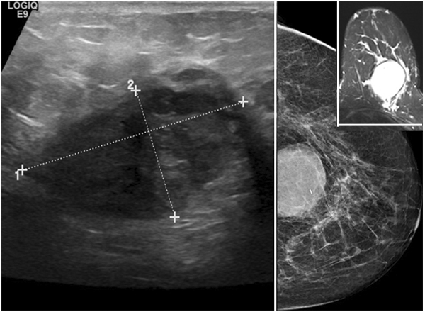
Histology
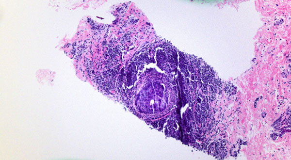
Core Biopsy
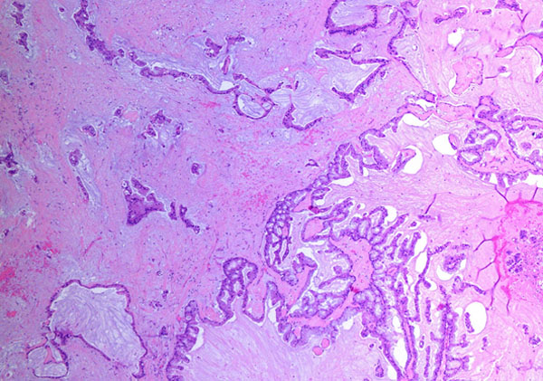
Cystically dilated spaces lined by papillary, tufted epithelium with basally oriented nuclei and intracytoplasmic mucin.
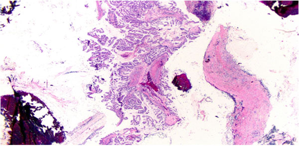
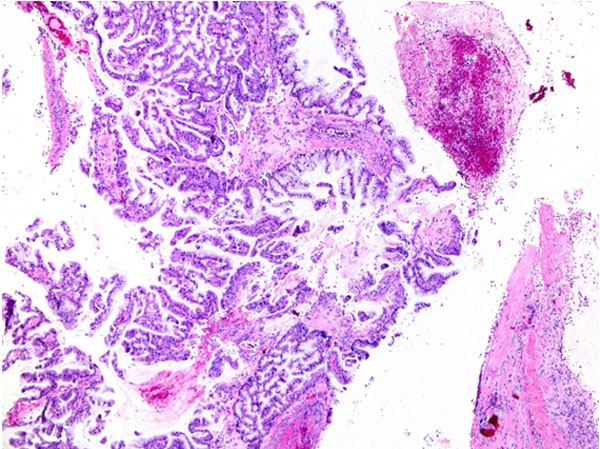
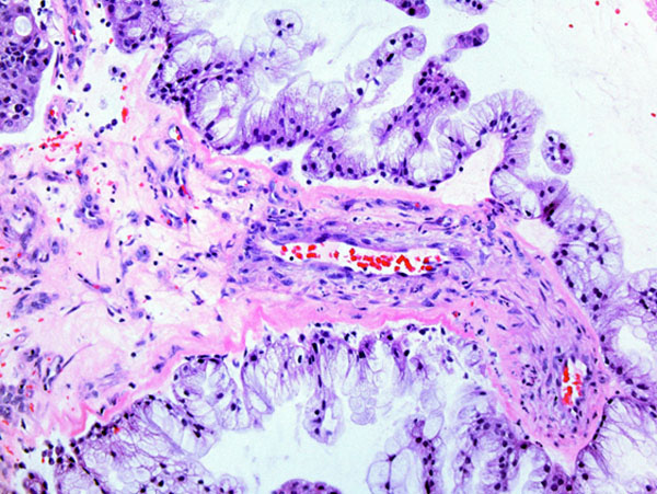
Variable nuclear atypia (from bland nuclei to moderate atypia) and cytoplasmic eosinophilia.
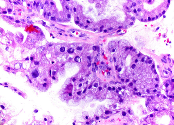
Focal ductal carcinoma in situ with periductal chronic inflammation.
Immunohistochemistry
CK7: Positive
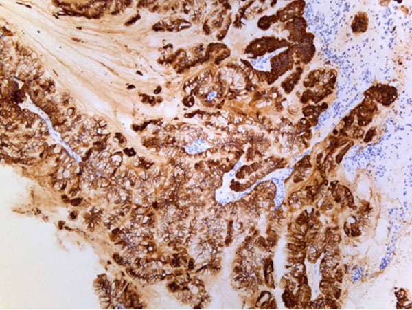
Mammoglobin: Positive
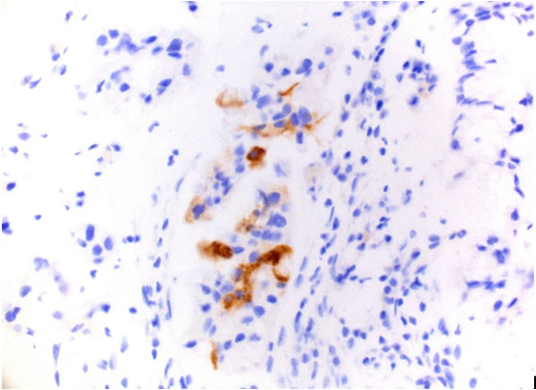
Negative stains: CK20, CDX2, PAX8, WT1, GCDFP15, GATA3, ER, PR, HER2
Excision
Left Breast Lumpectomy
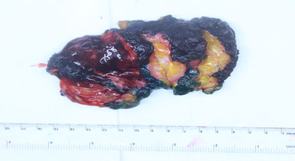
Gross: 6.2 x 4.4 x 4.0 cm cystic lesion, filled with opaque brown viscous fluid.
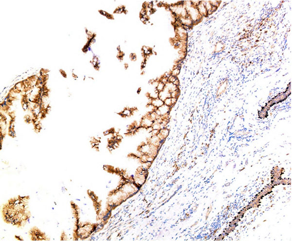
Intracellular and extracellular mucin
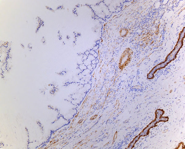
Myoepithelial markers negative: P63 and calponin
Final Diagnosis
Mucinous cystadenocarcinoma (MCA) of the breast.
Discussion
Mucinous cystadenocarcinoma of the breast (MCA) was first described by Koenig and Tavassoli in 1998 as a tumor composed of variably sized cystic spaces lined by neoplastic mucinous cells with bland, basally placed nuclei forming epithelial tufts and papillae. The interpretation of this tumor as an invasive carcinoma is confirmed by the absence of a myoepithelial cell layer on immunohistochemical staining. The 21 reported cases of MCA have shown varying degrees of cytologic atypia as well as varying degrees of mucin depletion and cytoplasmic eosinophilia. This characteristic tumor morphology mimics metastatic mucinous carcinomas of the ovary, pancreas and GI tract; thus, an IHC panel should be ordered including CK7+, CK20-, CDX2-, PAX8-, WT1-, MAMG+, GCDFP15+, GATA3+, ER-, PR-, and Her2-. The breast markers are highly specific but not sensitive; therefore, positive stains are contributory whereas negative stains are noncontributory. This entity should also not be confused with invasive mucinous carcinomas or signet ring cell carcinomas of the breast.
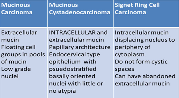
Invasive mucinous carcinoma contains extracellular mucin and signet ring cell carcinoma contains intracellular mucin, but MCA can be distinguished from these entities as it has intra- and extracellular mucin, a characteristic tufting pattern, and it lacks the displaced nuclear cytology of signet ring cells.
The reported cases of MCA have shown that this entity does not behave clinically as other ER-/PR-/HER2- tumors with a high KI67 index (5-70%). The likelihood of lymph node metastasis does not correlate with pathologic staging (15%), and no recurrences or distant metastases have been reported so far. Thus, MCA is considered to be an indolent behaving tumor requiring complete excision with sentinel lymph node dissection. The role of chemotherapy and radiation in these tumors is unknown.
For pathologic staging purposes, the entire tumor should be measured as the invasive tumor component. MCA type ductal carcinoma in situ has been reported, but if myoepithelial markers are negative, the invasive component will account for the greatest dimension of the mass such as in this case.
References
- Koenign C, Tavassoli F: Mucinous Cystadenocarcinoma of the Breast. The American Journal of Surgical Pathology Issue: Volume 22(6), June 1998, pp 698-703.
- Nektarios K et al: Mucinous Cystadenocarcinoma of the Breast: The Challenge of Diangosing a Rare Entity. Rare Tumors 2017; volume 9;7016.