
Contributed by Dimitrios Korentzelos, MD and Gabriela M Quiroga-Garza, MD
Clinical History
The patient is a woman in her sixties who presented in clinic for initial consultation for a 4.5 cm, enhancing, left renal mass identified on CT urogram as part of gross hematuria workup earlier this year. She had cystoscopy which was negative, and urine cytology which was also negative. A CT a/p scan showed a heterogeneously enhancing, left mid-pole renal mass, partially exophytic, measuring 4.5 x 4.2 x 4 cm, consistent with a renal cell carcinoma. No left hydroureteronephrosis was seen. The bladder was unremarkable. There were right-sided benign renal cysts as well as benign cysts in the liver. The patient denied recurrence of hematuria and weight loss. There was no family history of kidney cancer. Finally, there was no evidence of adenopathy or metastatic disease.
Gross Examination
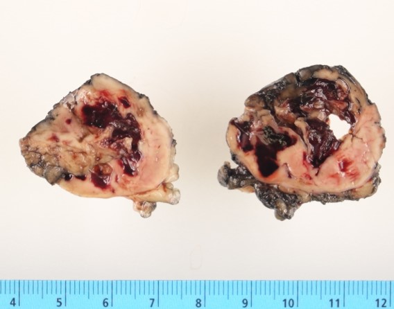
Gross examination revealed a 5.3 x 4.7 x 4.2 cm partial nephrectomy specimen, with an entirely exposed, disrupted and friable mass. The resection margins (capsular and parenchymal, as well as the area of disruption) were inked black. The cut surface revealed an ill-defined, disrupted, friable, tan-brown, focally hemorrhagic, lobulated and soft to rubbery mass (Figure 1).
Microscopic Examination
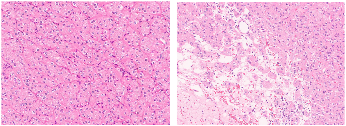
Histologic sections reveal an epithelioid neoplasm demonstrating predominantly solid growth of eosinophilic (oncocytic) cells with bland (low-grade) nuclei lacking nuclear atypia and irregularity. Perinuclear halos and occasional binucleate/multinucleate forms are present. Mitotic activity and tumor necrosis are not identified. Sharply delineated loose stromal areas with scattered clusters of neoplastic cells and focal osseous metaplasia are also noted. Finally, the neoplasm demonstrates scattered intratumoral lymphoid infiltrates (Figure 2).
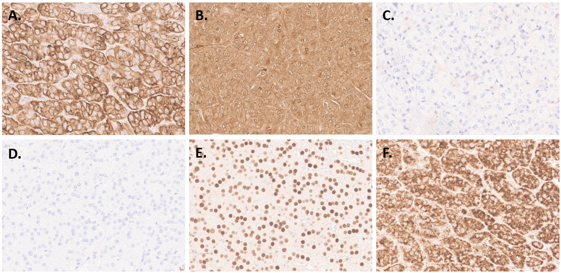
Immunohistochemical stains demonstrate the neoplastic cells are diffusely positive for CK7, parvalbumin, and GATA3, while they only show focal/weak c-kit expression. They have a very low Ki-67 proliferation index (<1%) and show preserved SDHB expression (Figure 3).
The overall findings support the morphologic impression of low-grade oncocytic tumor (LOT). However, the unlikely scenario of a chromophobe renal cell carcinoma with bland histologic features and an unusual c-kit expression pattern cannot be entirely ruled out, thus Oncomine next-generation sequencing (NGS) studies were performed to adequately characterize the tumor.
Molecular Studies
Next generation sequencing (NGS) studies performed on the formalin-fixed paraffin-embedded tissue demonstrate a TSC2 pathogenic/likely pathogenic (Tier 1/2) in-frame deletion with a variant allelic frequency (VAF) of 29%. No gene fusions or copy number alterations were detected, and the tumor was microsatellite stable (MSS) with a low tumor mutational burden (TMB) of 1 mutations/Mb (11 Percentile).
Diagnosis and Discussion