
Author: Kyle Hinz, MD
Mentor: Arivarasan D. Karunamurthy, MD
Clinical History
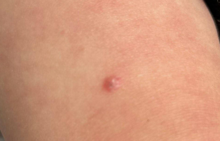
A toddler was brought into clinic for a concern of a pink nevus on her right anterior thigh. The lesion was initially flat and flesh colored. The lesion began to change, becoming raised, erythematous, and with central brownish pigmentation (Figure A). No pain, pruritus, or irritation was present. Additionally, no discharge or bleeding was present. A shave biopsy was sent for pathologic review.
Histologic Findings
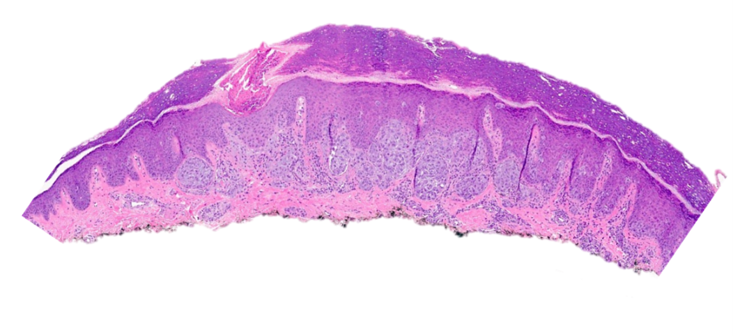
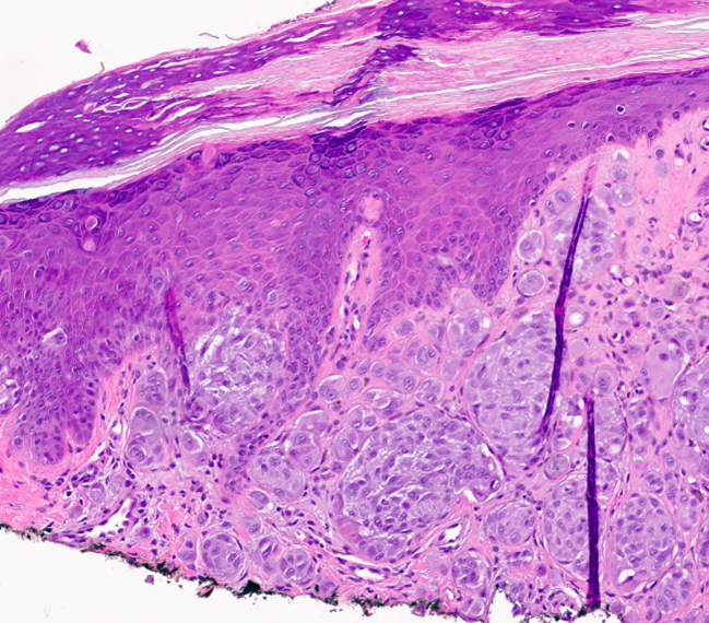
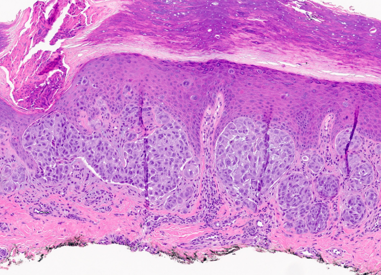
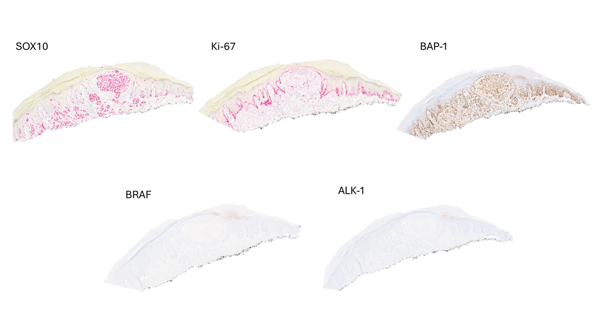
Shave biopsy of the right thigh showed a compound, predominantly intradermal proliferation of epithelioid melanocytes. The epidermis show small nests and single cells of epithelioid melanocytes are present at the tips of the dermal papillae. At the dermal-epidermal junction, rare Kamino bodies were noted. Within the dermis, nests of dyscohesive epithelioid melanocytes and single cell melanocytes limited to superficial dermis with enlarged nuclei, mild nuclear pleomorphism, occasional nucleoli, and amphophilic cytoplasm (Figure B-D). High grade features such as cytologic atypia, mitotic figures, ulceration, or expansile growth pattern was not identified. Immunohistochemistry showed lesional cells with retained p16, SOX10, and BAP1 nuclear stain. Β-catenin nuclear, BRAF VE, and ALK stains are negative (Figure E). Ki-67 shows a proliferation rate of <1%.
Molecular
FISH testing was ordered given the Spitz features and mild cytologic atypia. 4 probe FISH test targeting chromosome regions 6p24, 8q24, 9p21, and 11q was negative for copy number aberrations. Targeted DNA/RNA panel (Oncomine V3) Next Generation Sequencing demonstrated a kinase gene fusion, CCDC6/RET fusion.