
Authors: Katherine Killian, DO and Daniel Marker, MD, PhD
Clinical History
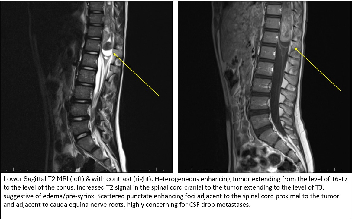
A young girl in her first decade presented for evaluation following a three-week history of insidious onset of severe back pain. Spine radiograph demonstrated a 20-degree angle of scoliosis with right vertebral pedicle asymmetry. A total spine MRI was ordered for further evaluation and revealed a heterogenous enhancing tumor extending from the level of T6-T7 to the level of the conus with evidence of drop metastasis at the cauda equina. Gross total resection was performed with T8-L1 laminoplasty and intramedullary tumor resection.
Microscopic Examination
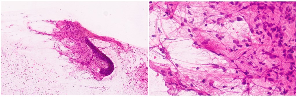
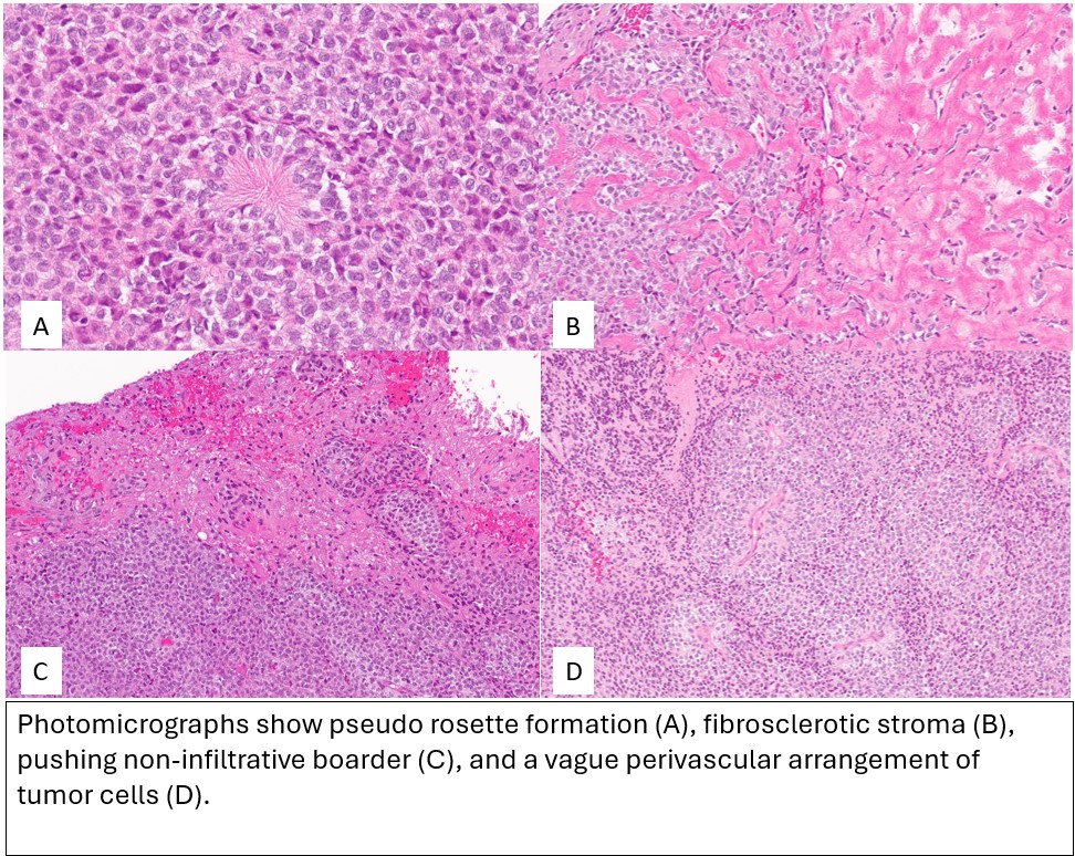
The smear preparation demonstrated neoplastic proliferation of small round blue cells with fine fibrillary processes. Histology revealed a fairly circumscribed primitive tumor composed of small round blue cells with pushing non-infiltrative borders. Focal perivascular structuring, rosette-like structures with central fibrillary processes, and areas of dense extracellular sclerosis are present. Mitotic figures are readily identified and there is multifocal tumor necrosis.
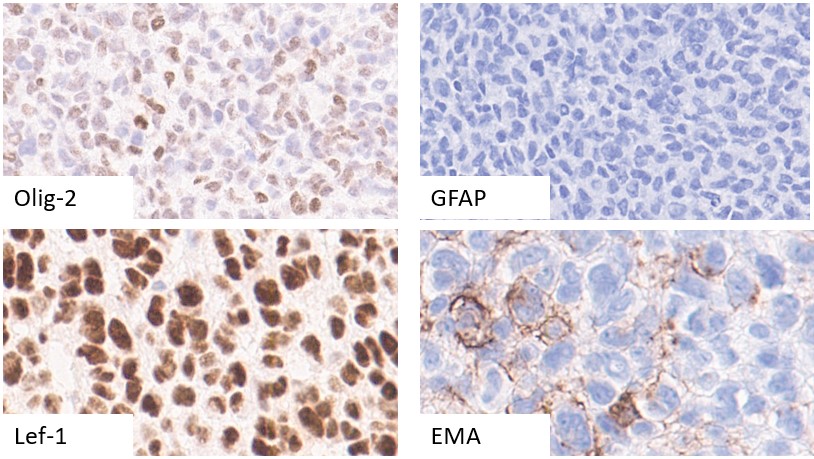
Immunohistochemical studies were performed. The tumor cells demonstrate diffuse strong nuclear positivity for Lef1, patchy positivity for OLIG-2, patchy membranous/cytoplasmic positivity for EMA, and are essentially negative for GFAP. Ki-67 is variable with areas approaching 25-30% within the tumor.
The morphologic features and immunophenotype were not specific for a diagnostic entity, but were suggestive of potential aggressive behavior. Next-generation sequencing and DNA methylation array classifier analysis were ordered. A Final Integrated Morphologic and Molecular Diagnosis was issued following their completion.