Authors: Haley Corbin, MD, Azfar Neyaz, MD, Rana Naous, MD
Clinical History/Pathological Findings
An elderly woman presented to orthopedic surgery musculoskeletal oncology with a right paraspinal soft tissue mass first noticed a few months ago. She was not sure if it had grown in size but it had become uncomfortable when sitting in a chair. She denied any recent weight loss, fevers, chills, or other lumps. On exam, there was a palpable soft tissue mass on the mid-back in the right paraspinal area which was tender to palpation with no erythema or other overlying skin changes.
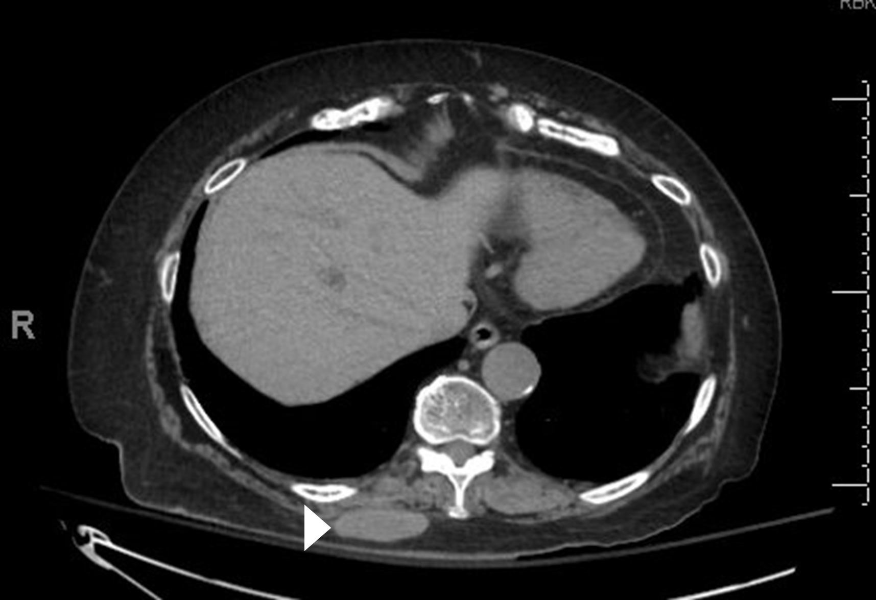
CT abdomen and pelvis without contrast showed an enlarged, well-marginated, partially calcified nodule in the deep subcutaneous soft tissues and subcutaneous fat in the right posterior lower thorax measuring 5.2 x 4.5 x 1.9 cm (Figure 1). This had gradually increased in size since 2022 when it measured up to 2.4 cm. Chest CT showed lung metastases.
On biopsy of the lesion, histologic sections demonstrated nests and sheets of predominantly round to focally short spindle cells, with intervening areas of fibrosis and focal necrosis. The round tumor cells showed high N:C ratio, prominent nuclear irregularity, conspicuous nucleoli and scant cytoplasm. Frequent mitotic figures were noted (up to 8 per 10 high-power fields [HPF]) in the areas with round cell morphology. The tumor cells were positive for vimentin, CD34, STAT6, synaptophysin (focal), and SMA (focal), while negative for CK7, TTF-1, CK20, CK19, chromogranin, Melan-A, S100, HMB45, CD10, LCA, desmin, p40, desmoglein3, WT1 and SOX10. Cam5.2 and AE1/3 demonstrated very rare staining. Ki-67 highlighted approximately 30% of tumor cells in hot spots. TLE1 showed weak nonspecific staining. CD34 shows loss of expression in the round cell component.
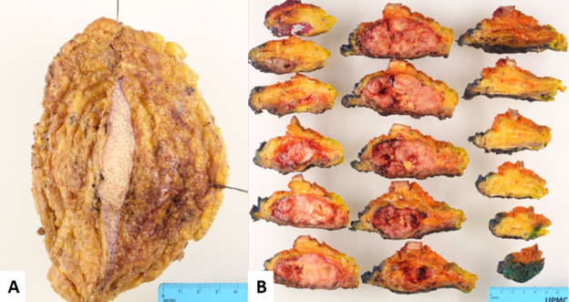
The patient underwent palliative resection of the mass followed by systemic therapy. The resection specimen was received by pathology as an excision of adipose tissue, skin, and skeletal muscle (Figure 2, A) which was sectioned to reveal a 6.8 x 6.7 x 3.5 cm tan-pink, glistening mass with mainly well demarcated and thinly encapsulated borders (Figure 2, B). The mass was tan-white and rubbery with fleshy areas of hemorrhage and areas of yellow, friable necrosis.
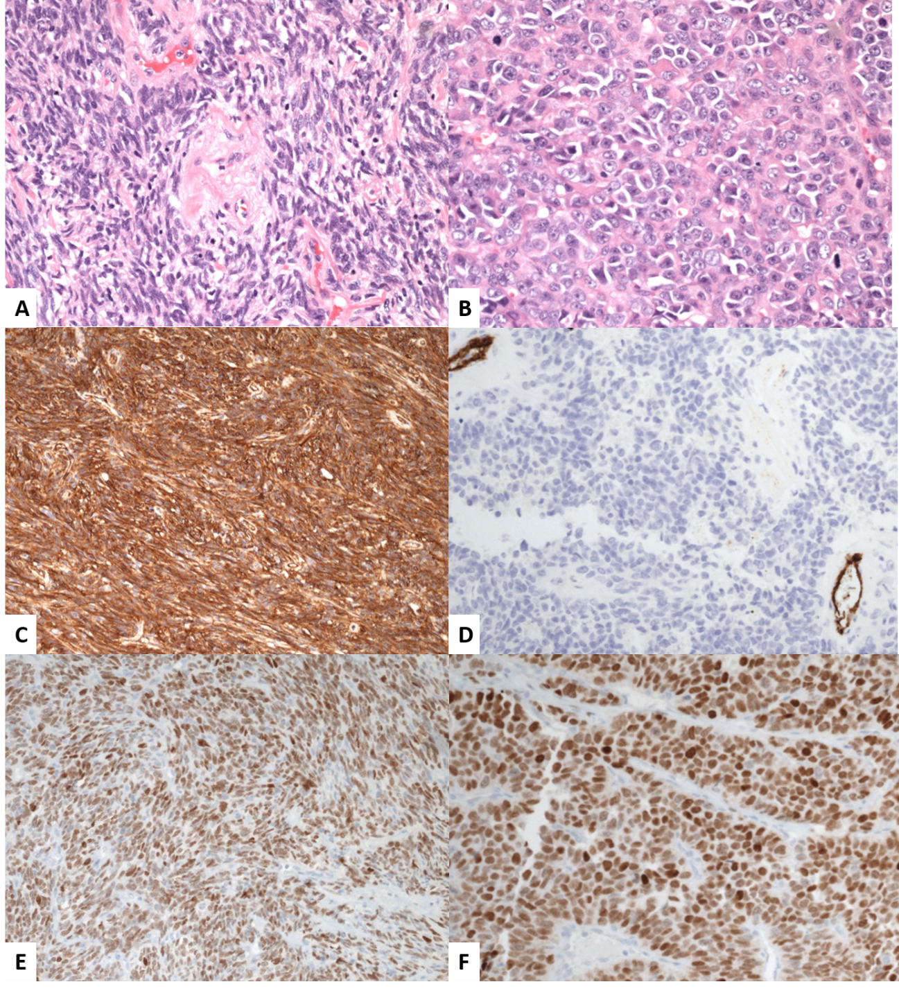
Microscopic examination of the tumor resection specimen revealed sheets and nests comprised predominantly of round cells with vesicular chromatin, prominent nucleoli, and high N:C ratio (Figure 3, B). Focal spindled cell areas comprised a minor component of the tumor with cells arranged in short and long fascicles with perivascular hyalinization (Figure 3, A). Hemorrhage and osteoclast-type giant cells were noted focally. Mitotic figures reached 60 per 10 HPF and necrosis was estimated at 10%.
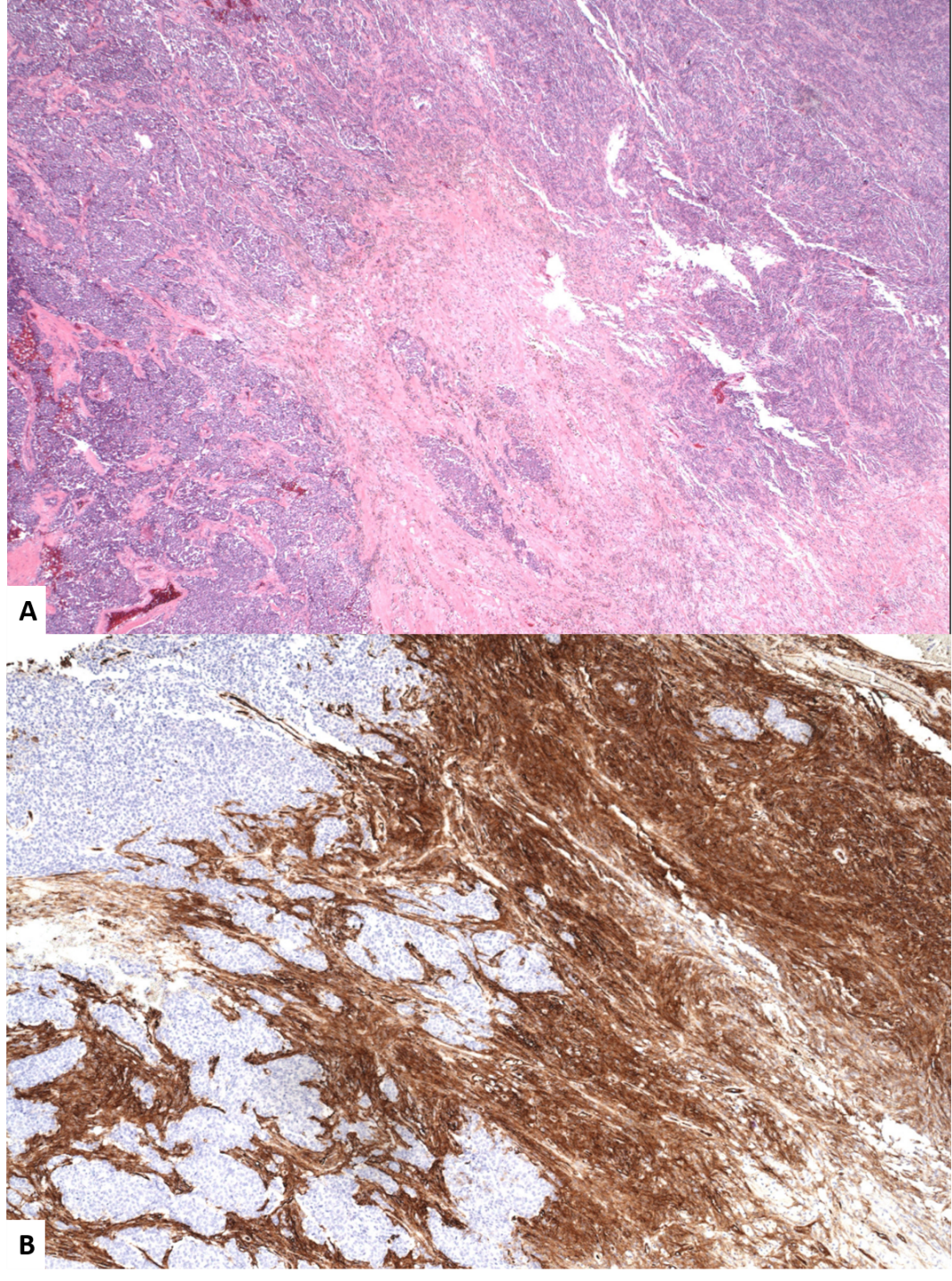
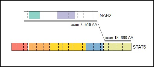
Immunohistochemical studies showed that the tumor cells were positive for STAT6 with variable intensity (Figure 3, E and F). and focally positive for SMMHC. The spindle cell component was positive for CD34 (Figure 3, C) with loss in the round cell high-grade component (Figure 3, D; Figure 4). SMA and AE1/AE3 showed patchy positivity. The tumor cells were negative for S100, SOX10, HMB45, Melan-A, desmin, myogenin, ERG, ALK1, MyoD1, WT1, NUT, caldesmon, and MUC4. H3K27me3 and INI-1 were retained in the tumor cells. FISH for EWSR1 gene rearrangement showed a complex signal pattern. FISH for SYT and TFE3 gene rearrangements and MDM2 gene amplification were negative. RNA seq showed NAB2::STAT6 gene fusion (Figure 5), and Oncomine molecular testing revealed TERT and TP53 gene mutations and multiple copy number alterations.