
Submitted by Seemaab Ali, MD, PhD, and Robert L. Peel, MD
Clinical History
The patient is a woman in her 60s with a one-year history of chronic sinusitis with nasal congestion and loss of smell, as well as a four-month history of right eye proptosis and blurred vision with diplopia and chronic tearing from the right eye. She experienced no benefit from antibiotics or steroids, and imaging of the sinuses and orbit was performed.
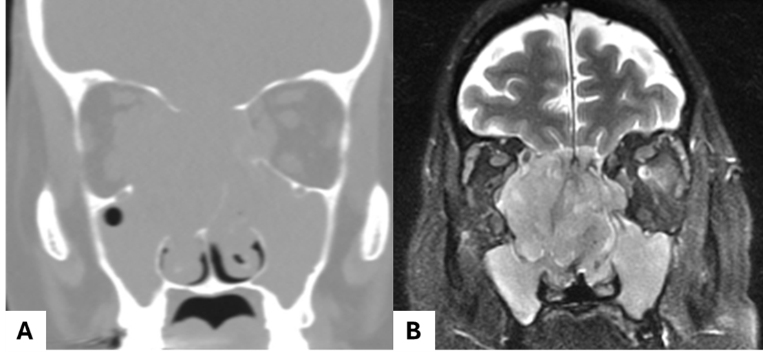
CT imaging demonstrated complete opacification of the sinuses, with expansion of the orbits and orbital and skull base bony erosion. Anterior rhinoscopy and nasal endoscopy showed only edematous mucosa and did not reveal an obvious tumor. MRI imaging showed a large bilateral nasal cavity mass measuring approximately 5 cm with extensive involvement of the sinuses, orbital floors, and intracranial and right orbital extension. Given the aggressive appearance of the tumor, biopsies were performed.
Histologic Findings
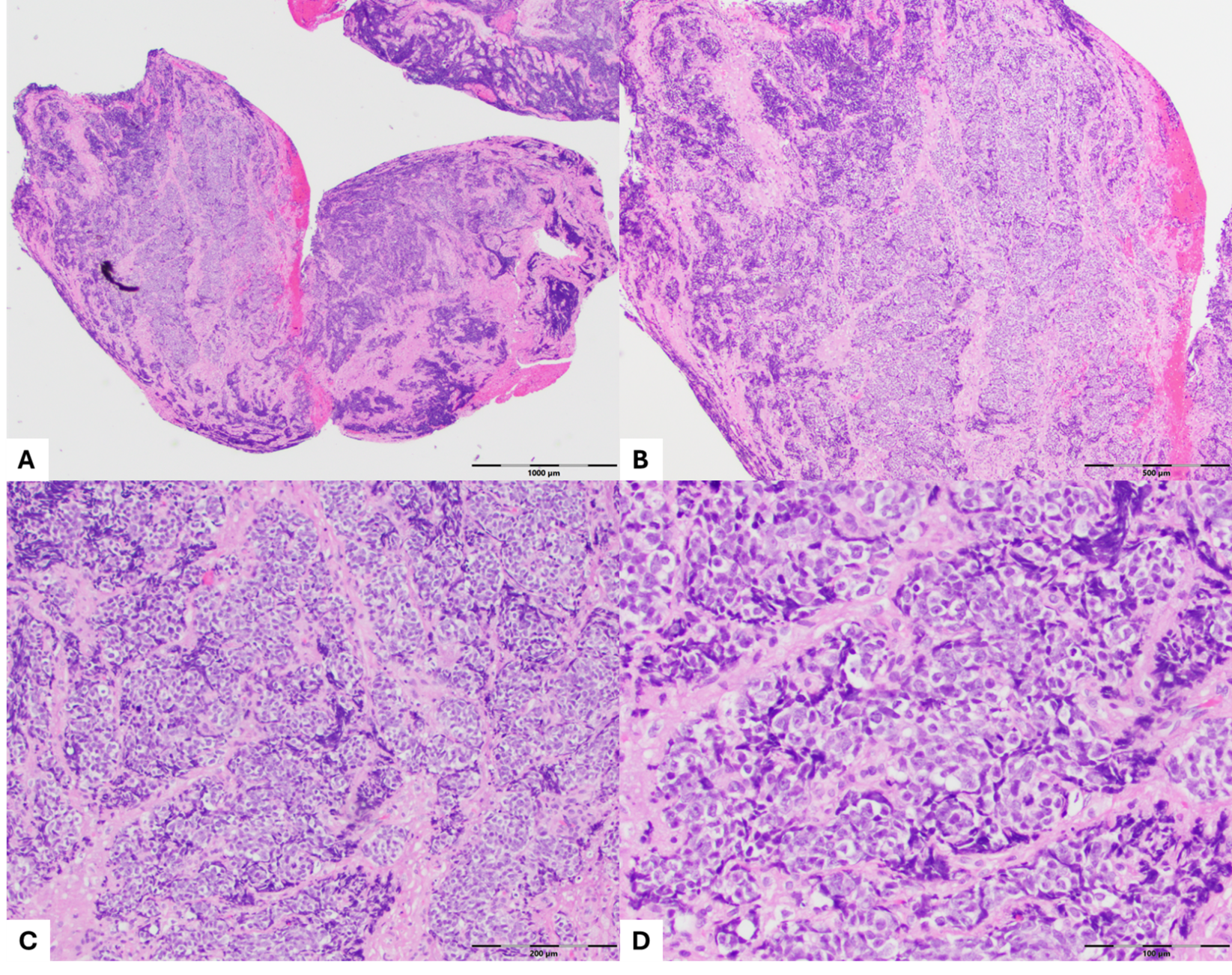
Histologic sections demonstrate an invasive neoplasm with vaguely nested to sheet-like infiltrative growth pattern, consisting of rounded, undifferentiated-appearing cells. The cells exhibit large, rounded and centrally located hyperchromatic nuclei, prominent and eosinophilic nucleoli, high nuclear:cytoplasmic ratios, and cytoplasmic clearing. Crush artifact is present extensively (Figure 2).
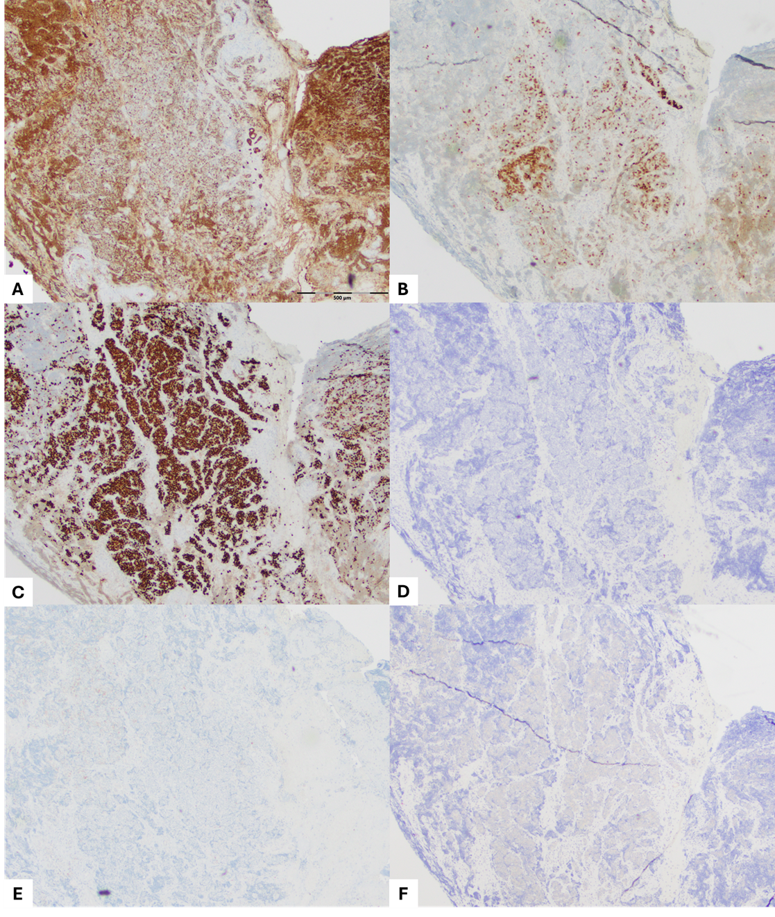
Immunohistochemistry shows positive staining for a pankeratin and somewhat patchy staining for p16. p53 demonstrates a mutant pattern. The tumor is negative for p40, Melan A, synaptophysin, and NUT. Retinoblastoma, SMARCA2, BRG1 (SMARCA4), INI1 (SMARCB1), and ARID1A are retained (Figure 3).
Oncomine molecular testing is ordered to support the final diagnosis.