Authors: Matthew Chambers M.D., Ph.D., Sheldon Bastacky, M.D.
Clinical History
A man in his seventh decade of life presents with a unilateral testicular mass, approximately 3-4 cm, which is clinically concerning for malignancy. Serum markers are drawn, and both AFP and beta-hCG are within normal limits. A radical orchiectomy is performed.
Gross Examination
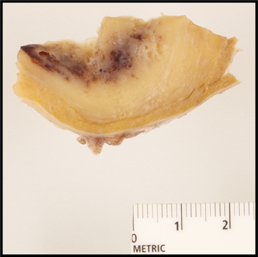
A well-circumscribed, 5 cm lesion with tan solid areas, small cystic spaces, and areas with appearance of hemorrhage (40% of the lesion) are seen. Minimal residual testicular parenchyma is present, and the lesion appears to be grossly confined to the testis.
Histologic and Immunohistochemical Ancillary Studies
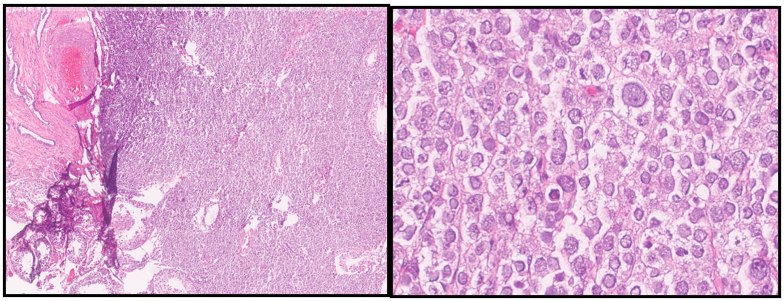
Histologic sections of the lesion are then assessed microscopically (see Figure 2).
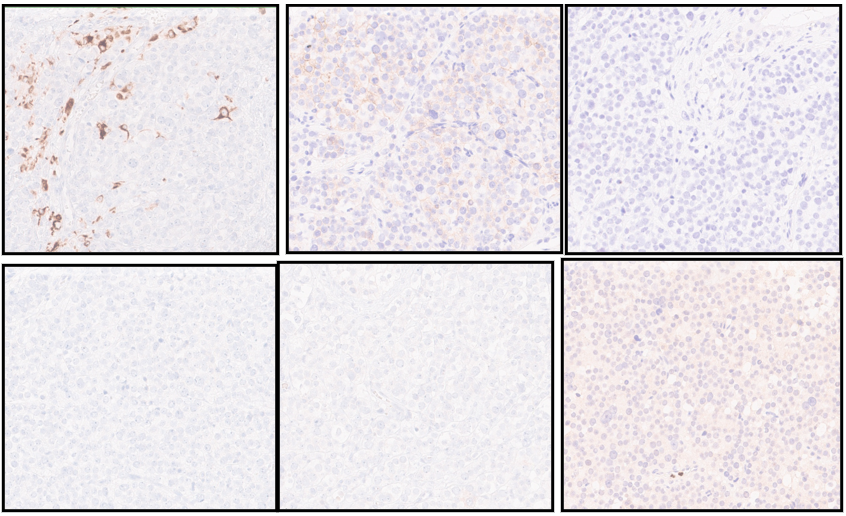
Additional ancillary immunohistochemical stains are performed (pan-keratin, c-kit (CD117), OCT3/4, CD30, AFP; see Figure 3). The tumor shows only very rare immunoreactivity for pan-keratin (<1%), patchy and weak immunoreactivity for c-kit (CD117), and is negative for OCT3/4, CD30, AFP and OCT2.
Cytogenetic
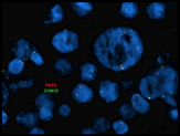
Given the above immunohistochemical findings, follow-up cytogenetic studies were performed to assess for the presence of chromosome arm 12p via fluorescence in situ hybridization (FISH) studies; however, assessment for the presence of 9p was not available on-site. The results showed that 12p was over-represented in the examined cells, which can be seen in the presence of an isochrome 12p, 12p duplication or chromosome 12 hyperploidy.