Authors: Simmi Patel, M.D. and Daniel F. Marker, M.D., PhD
Clinical History
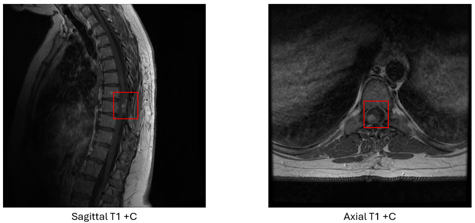
A female in her 6th decade presented to an outside institution 3 years ago with initial symptoms of difficulties walking and paresthesia. MRI showed an intradural extramedullary mass with some contrast enhancement in the thoracic spinal cord. The mass was initially resected, and a diagnosis was issued. The tumor recurred 3 years later and was re-resected.
Microscopic Examination
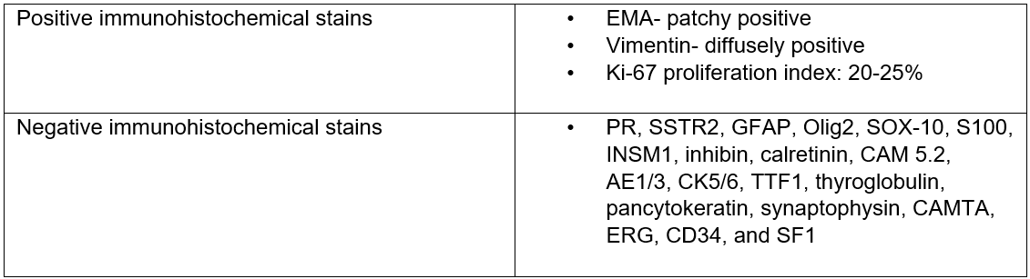
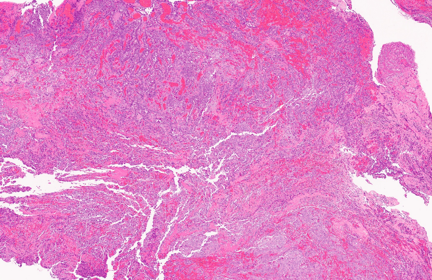
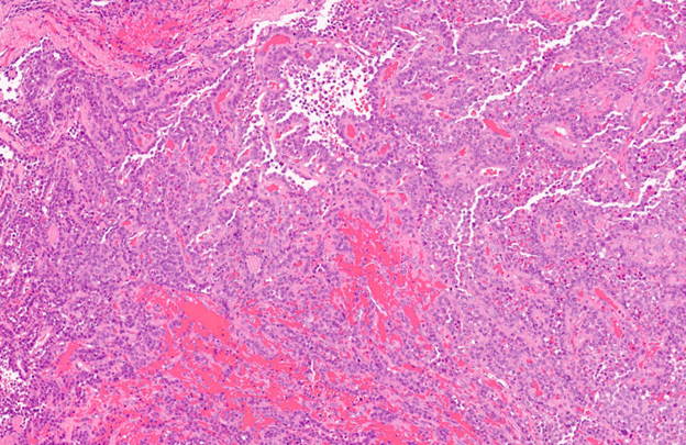
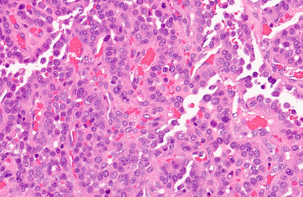
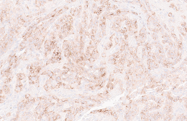
Initial histologic sections showed a neoplasm composed of atypical pleomorphic cells arranged in a papillary/pseudoangiomatous growth pattern. An extensive immunohistochemical stain panel was conducted seen in table 1. The tumor was mostly positive for EMA (patchy) and vimentin (diffuse). The Ki-67 proliferation index was 20-25%.
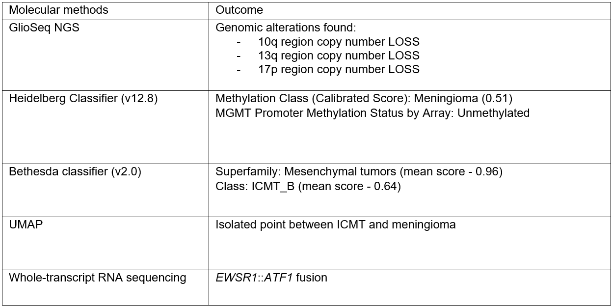
Molecular Studies