
Contributed By: Daisy Wu, MD, Diana M. Bell, MD
Clinical History
A man in his seventh decade of life presents with an upper lip lesion present for the past 10 years. Throughout the past decade, he has been noting slight growth of the lesion and occasionally accidentally cutting over his skin over the lesion during shaving.
Gross Findings:
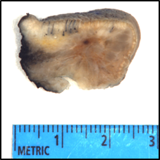
Lip resection demonstrates a 2.0 x 1.1 x 0.5 cm raised irregular ill-defined subcutaneous lesion under tan-gray and tan-pink mucosa (see Fig. 1).
Histopathologic Features:
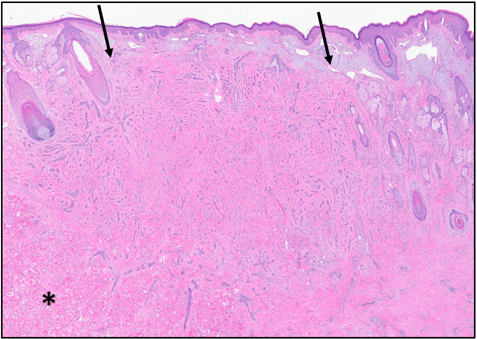
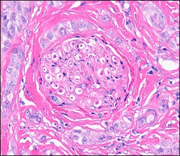
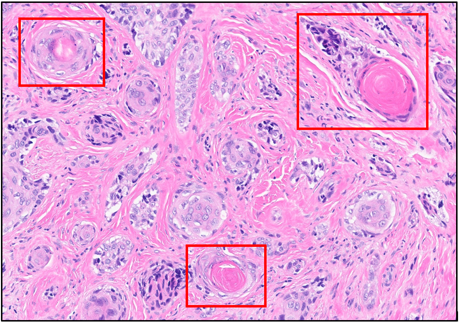
Histological analysis of lesion demonstrates subcutaneous sclerosing basaloid infiltrative solid nests, strands and columns of small monomorphous epithelial cells and ductal structures composed of two layers of small cuboidal cells obliterating adnexal structures within the region (Fig. 2). These lesional cells also demonstrate infiltration into adjacent skeletal muscle as well as perineural invasion (Fig. 3). The tumoral cells forming ducts also occasionally demonstrate eosinophilic concentric secretions consistent with keratin suggestive of trichilemmal differentiation.
Whole slide images for this case can be viewed at (use theViewOnline links): Digital Slide Viewer: MAC
Immunoperoxidase studies was performed on the lesion including p40, CK5/6, CK7, CK34, CEA-M, SOX-10 and Ki-67. Tumor cells were diffusely positive for p40, CK5/6, CK34, patchy positive for CK7, negative for CEA-M and SOX-10 and a Ki-67 proliferative rate around 3%.
Molecular findings:
UPMC Oncomine Test demonstrated an ARID1A p.Q766* mutation, ROS1 p.P1941Lfs*5 variant of uncertain significance and RAD51D p.Q177L variant of uncertain significance. The tumor mutation burden was at the 36 percentile.
What is the diagnosis?