Authors: Jacob Jerome, MD and Jocelyn Boccella, DO
Year of Publication: 2025
Clinical History
A woman in her 50s had a 1.2 cm irregular mass with architectural distortion and spiculation discovered on her screening mammogram. Follow-up ultrasound demonstrated a 1.4 cm irregular hypoechoic mass with angular margins. Ultrasound-guided core biopsy was performed.
Microscopic Examination
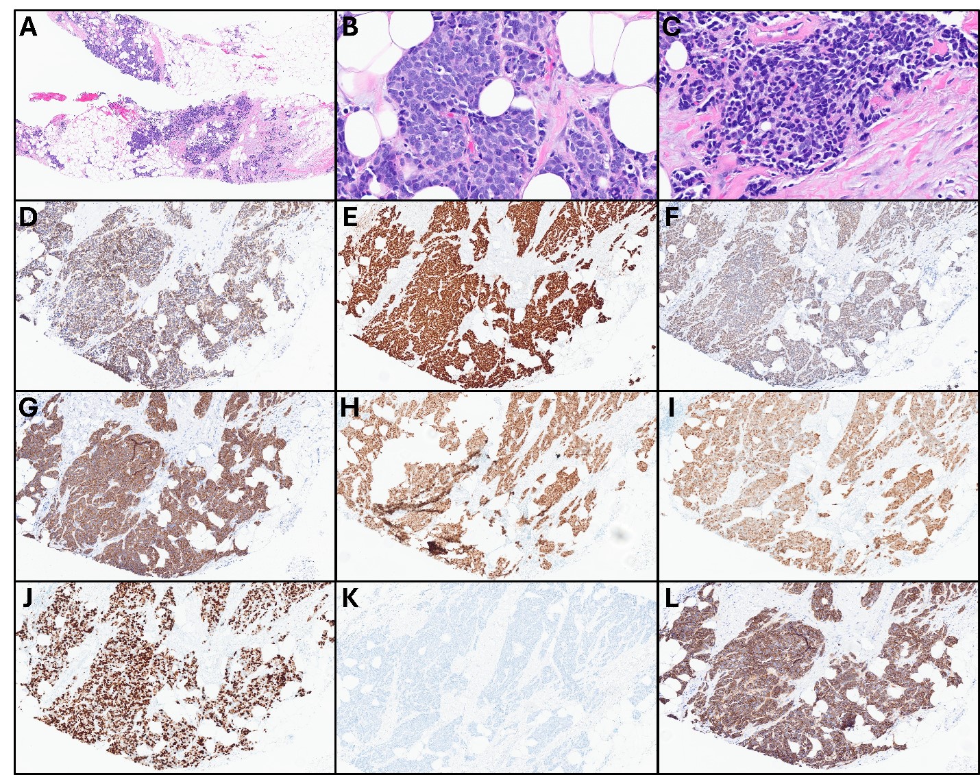
Biopsy cores demonstrated infiltrative nests of malignant-appearing cells (Figure 1A). The cells showed very high nuclear:cytoplasmic ratios with scant cytoplasm, finely granular chromatin, and inconspicuous nucleoli (Figure 1B). Mitotic figures, nuclear molding, and apoptotic debris were prominent features (Figure 1C). Immunohistochemical stains demonstrated the cells were positive for AE1/3 (Figure 1D), GATA3 (Figure 1E), TRPS1 (Figure 1F), synaptophysin (Figure 1G), chromogranin (Figure 1H), estrogen receptor (Figure 1I), and progesterone receptor (Figure 1J). HER2 was negative (Figure 1K). Ki-67 was very high, at approximately 90% (Figure 1L).