Authors: Bethany Batson, MD, PhD and Jing Yu, MD, PhD
Date of Publication: October 1, 2025
Case Presentation
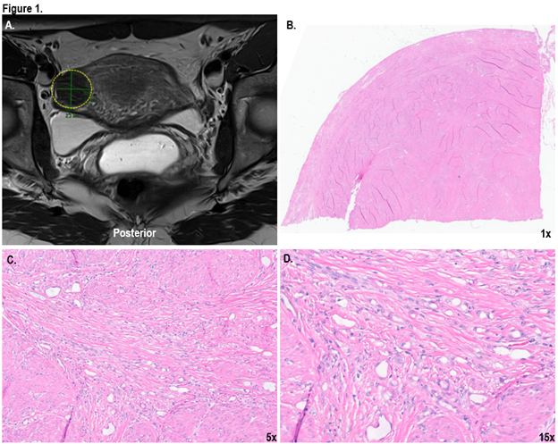
The patient is a 30-year-old female with a past history notable for cervical adenocarcinoma in situ and cervical intraepithelial neoplasia 1 and 2 in the setting of high risk HPV positivity which were previously resected by cold knife cone. Several years following treatment, atypical glandular cells were again identified on PAP screening. Subsequent endocervical curettage and endometrial biopsy yielded benign findings. Due to recurrent cytologic abnormalities, the patient elected to undergo a hysterectomy with bilateral salpingectomy as definitive treatment. Preoperative pelvic magnetic resonance imaging was notable for a 2.5 cm well-circumscribed T2 hypointense subserosal mass within the right anterior uterus which was radiologically consistent with a hypercellular subserosal uterine fibroid (Figure 1A). The hysterectomy and bilateral salpingectomy were performed without complication.
Gross examination of the uterus revealed a well circumscribed, homogenous white and whorled myometrial nodule comprising 10% of the total myometrium volume. With the exception of a small areas of puckered scar on the cervix, no other atypical macroscopic findings were noted. Given the patient’s history of cervical dysplasia, the entire cervix was submitted for microscopic examination in addition to representative sections of the myometrial nodule. Histologic sections of myometrial nodule revealed prominent intersecting bands of apparent smooth muscle with small simple gland-like spaces forming bridging strands between the smooth muscle fascicles, simulating infiltrative tumor cells. The gland-like spaces were lined by flat to cuboidal epithelioid cells without atypia (Figure 1B-D). To further characterize the nodule, a panel of immunohistochemical stains was performed. The gland-like and cystic proliferation was positive for mesothelial markers including calretinin, WT1 and D2-40, and was negative for epithelial marker MOC31. Given the morphologic and immunophenotypic profile of the tumor, the final diagnosis of adenomatoid tumor of the uterus was made.