Authors: Rachel Vanderschelden, MD and Tatiana Villatoro, MD
Date of Publication: November 1, 2025
Clinical History
A woman in her 60’s underwent MRI-guided core biopsy of the breast following abnormal imaging findings.
Microscopic Findings
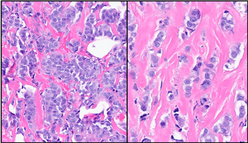
Core biopsy revealed an Invasive Mammary Carcinoma displaying predominantly ductal features, with focal areas exhibiting lobular features. The provisional tumor assignment was Nottingham Grade II, based on a total score of 7/9 (tubule formation: 3, nuclear pleomorphism: 2, mitotic rate: 2).
The carcinoma showed sparse stromal tumor infiltrating lymphocytes (sTILs) measured at 1%.
Associated findings within the biopsy specimen included ductal carcinoma in situ (DCIS) nuclear grade 2, solid type with comedonecrosis and calcifications. Additional benign and proliferative lesions included atypical ductal hyperplasia (ADH), fibrocystic changes, usual ductal hyperplasia, sclerosing adenosis, apocrine metaplasia, and benign luminal calcifications.
Immunohistochemistry
ER: Strongly positive (H-score 260)
PR: Strongly positive (H-score 240)
HER2: equivocal by IHC (2+); amplified by FISH
Ki-67: Very high (80%).
E-cadherin: Predominantly membranous staining, with focal attenuation.
p120: Predominantly membranous staining, with focal cytoplasmic localization.