Author: Zobash Noor, MD
Mentor: Arivarasan Karunamurthy, MD
Date of Publication: February 1, 2026
Clinical History
The patient is a 63-year-old male with a dermatologic history significant for multiple moderately dysplastic nevi previously excised from the left mid back and left upper back between 2008 and 2015, without evidence of recurrence, and no personal history of skin cancer. He has a family history notable for malignant melanoma in his mother. The patient presented for evaluation and biopsy of a clinically atypical fleshy papule on the right lateral neck. The patient underwent tangential shave biopsies of the lesion, and the specimen was submitted to dermatopathology for histologic evaluation.
Gross Description
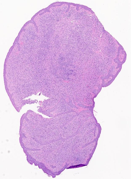
Gross examination of the right lateral neck shave biopsy specimen revealed a yellow-tan, pedunculated soft tissue fragment measuring 0.3 × 0.3 × 0.7 cm. The specimen was inked to designate the peripheral margins and submitted entirely for histologic evaluation.
Microscopic Examination
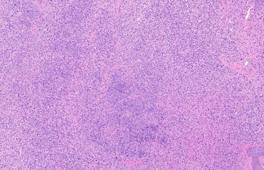
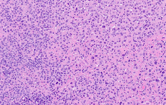
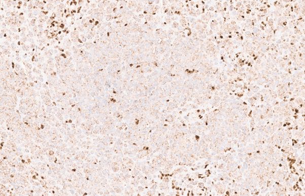
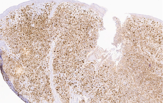
Histologic evaluation of the right lateral neck shave biopsy demonstrated a compound melanocytic neoplasm involving the epidermal junction, superficial dermis, and mid dermis. The lesion was composed predominantly of epithelioid melanocytes arranged in nests and sheets. The melanocytes exhibited abundant amphophilic cytoplasm and enlarged vesicular nuclei with prominent nucleoli. Mild cytologic atypia was present overall, with occasional lesional cells showing moderate nuclear enlargement, nuclear pleomorphism, and prominent nucleoli. A mild chronic inflammatory infiltrate was noted within the dermis. No tumor necrosis was identified, and mitotic activity was low.
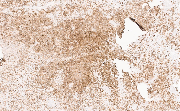
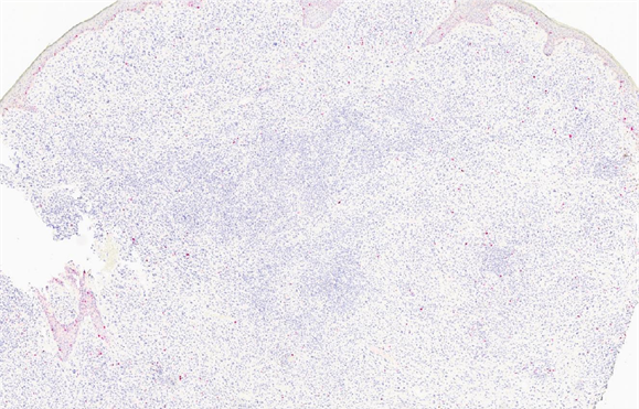
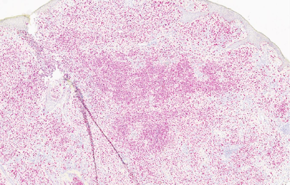
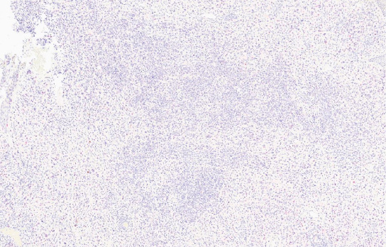
Immunohistochemical studies showed diffuse expression of SOX10 with a distribution of melanocytes consistent with the lesion. PRAME staining was negative. BRAF immunostaining was positive. Loss of nuclear BAP1 expression was identified in the lesional cells, with retained expression in background non-neoplastic cells, supporting BAP1 inactivation. Ki-67 highlighted a low proliferative index of approximately 1–2% of lesional cells, and p16 expression was preserved. The lesion extended to the deep biopsy margin. Overall, the histomorphologic and immunophenotypic features support a diagnosis of BAP1-inactivated melanocytoma with mild atypical features.