Authors: Tiarra Price, DO and Dimitrios Korentzelos, MD
Date of Publication: March 1, 2026
Clinical History
A woman in her 60’s with history of liver transplant, NASH, cirrhosis, hypertension, hyperlipidemia, sarcoidosis and chronic kidney disease presented with fever, flank pain and dysuria. Imaging showed a nearly 4 cm renal fluid collection concerning for abscess. The collection was drained, and cultures subsequently grew Escherichia coli. The patient was discharged on antibiotics.
The abscess persisted in the following months despite numerous interventions. Although the patient was feeling better, follow up imaging demonstrated a 7.6 cm heterogenous renal mass showing extension beyond the renal capsule extending within the peri-renal space laterally abutting the posterior inferior margin of the liver. The decision was made to pursue biopsy and subsequently radical nephrectomy.
Gross and Microscopic Findings
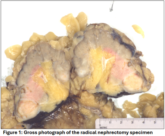
Gross examination of the radical nephrectomy specimen revealed multifocal, ill-defined irregularities with focal necrosis.
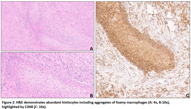
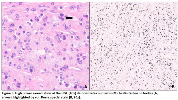
Microscopy revealed sheets of histiocytes (highlighted by CD68) and intracytoplasmic basophilic inclusions (Michaelis-Gutmann bodies, highlighted by a von Kossa special stain) with acute and chronic inflammation, foamy macrophage aggregates, fibrosis, and focal necrosis (Figures 2 and 3). A pankeratin immunostain was negative.