
Authors: Timothy Ramseyer, MD, MPH; Kyle Hinz, MD, MS; Samer Khader, MD
Date of Publication: March 1, 2026
Clinical History
A female in her 30s with a history of ALK-positive anaplastic large cell lymphoma in remission following chemotherapy presented for a follow-up due to abnormal laboratory values. The patient had intermittent abdominal pain, mucositis, and elevated INR, AST, and ALT liver function tests for several months, so imaging was ordered which identified a liver lesion. Shortly thereafter, the patient underwent ultrasound guided fine needle aspiration of the liver lesion.
Imaging
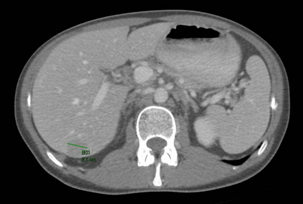
An axial post contrast CT abdomen shows a 2.1 cm subcapsular hypovascular liver lesion in segment 7 of the right hepatic lobe. No additional lesions or masses were identified.
Microscopic Examination
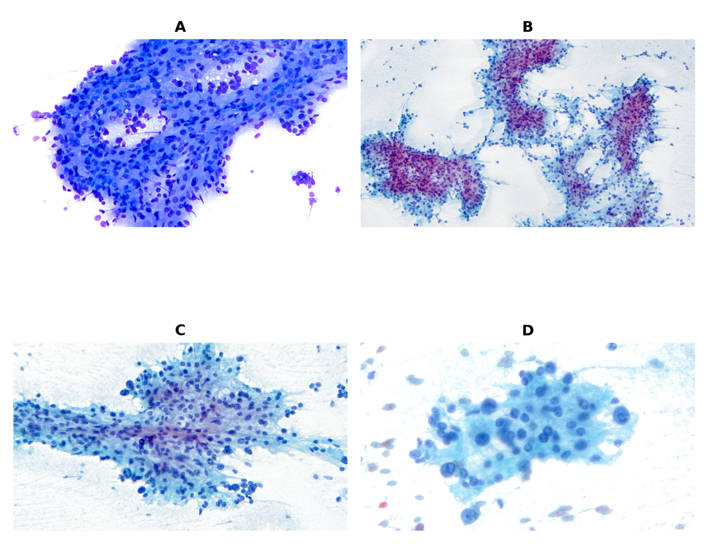
The Diff Quick (A) and Papanicolaou-(B-D) stained aspirate smear slides are cellular, showing loosely cohesive clusters, sheets, and singly dispersed cells, sometimes with perivascular-like arrangement. The morphology of the tumor cells was variable with round, polygonal, epithelioid, and occasional spindled morphology. The cell block (second figure) was cellular with the sample epithelioid, polygonal, and spindled cells. The neoplastic cells have abundant eosinophilic, finely vacuolated cytoplasm with nuclei exhibiting mild to moderate atypia and prominent nucleoli. No overt mitotic figures or tumor necrosis are identified. Immunostains performed on the cell block material showed the lesional cells to be negative for CD30, ALK, and Granzyme B. The lesional cells were positive for SMA, Caldesmon, HMB-45, and Melan-A.

Diagnosis and Discussion