
Authors: Claire Wilcox, MD, and Mahmoud Aarabi, MD, PhD
Date of Publication: March 1, 2026
Clinical History
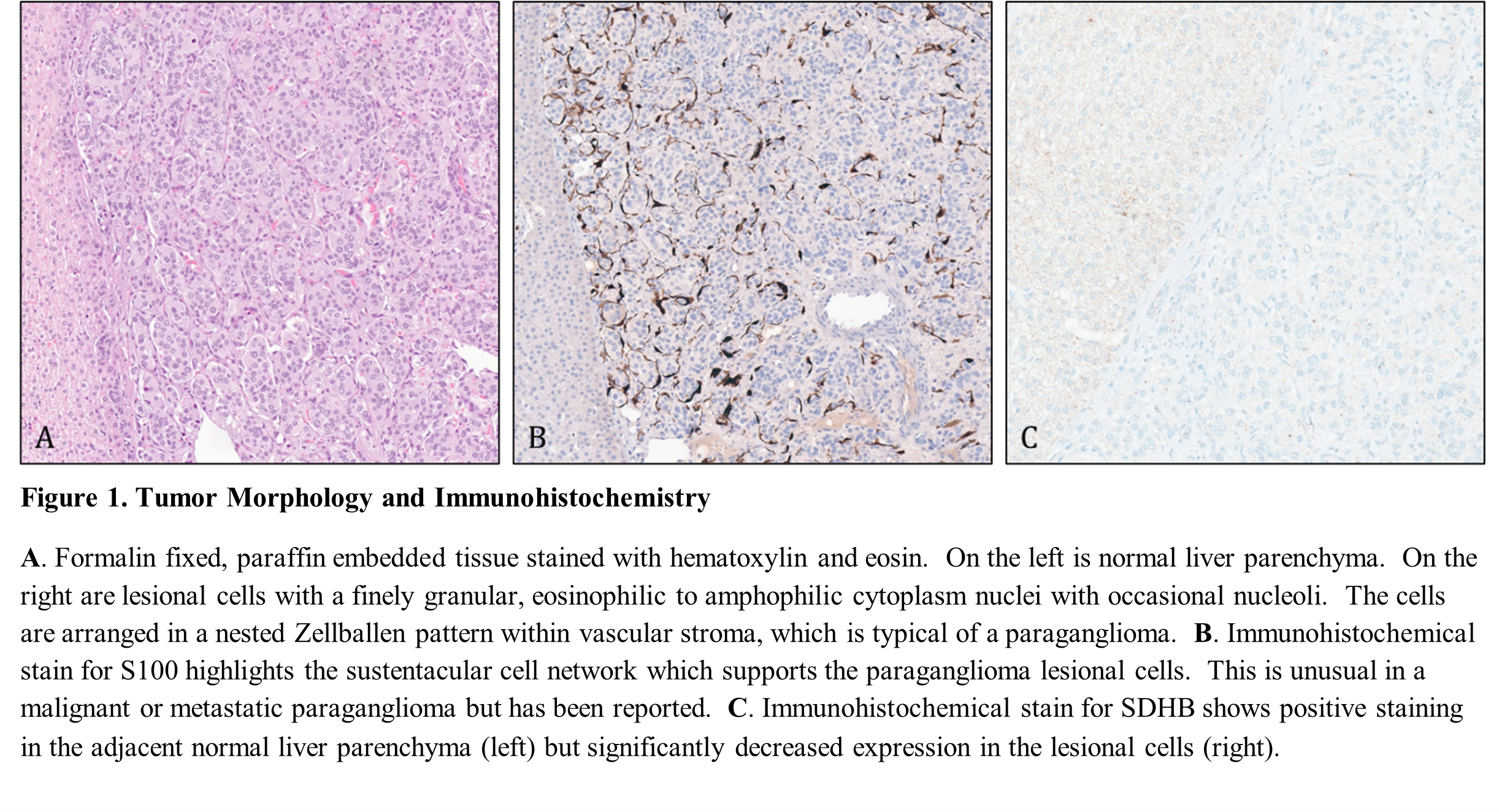
A female patient in her teens presented with multiple months of back pain, initially managed conservatively. Due to lack of improvement, MRI of the spine was performed and revealed a 12 cm suprarenal mass along with additional small masses in the liver and lungs. Elevated total urine metanephrines, normetanephrines, and total plasma metanephrines were detected in the laboratory workup. Liver biopsy confirmed the presence of paraganglioma (Figure 1-A). The multiple lesions, including the largest mass in the area of the adrenal gland, were deemed to represent either metastases of pheochromocytoma or multiple primary paragangliomas. The presence of a retained S100-positive sustentacular cell network in the liver lesion (Figure 1-B) suggested a synchronous multifocal primary tumor; metastases typically lack sustentacular cell networks, although rare metastases may have preserved sustentacular cells (Unger et al., 1991). Given that primary paragangliomas have been reported to arise in any tissue with sympathetic or parasympathetic ganglia (effectively any tissue except lymph nodes or bone) (Mete et al., 2022), the smaller lesions could not be definitively classified as metastases or multiple primary lesions. SDHB immunohistochemical stain was markedly decreased, with rare punctate cytoplasmic staining (Figure 1-C). It was suggested that genetic testing be performed for possible driving pathogenic variants in multiple genes including SDHB and SDHD.
Molecular Testing Results
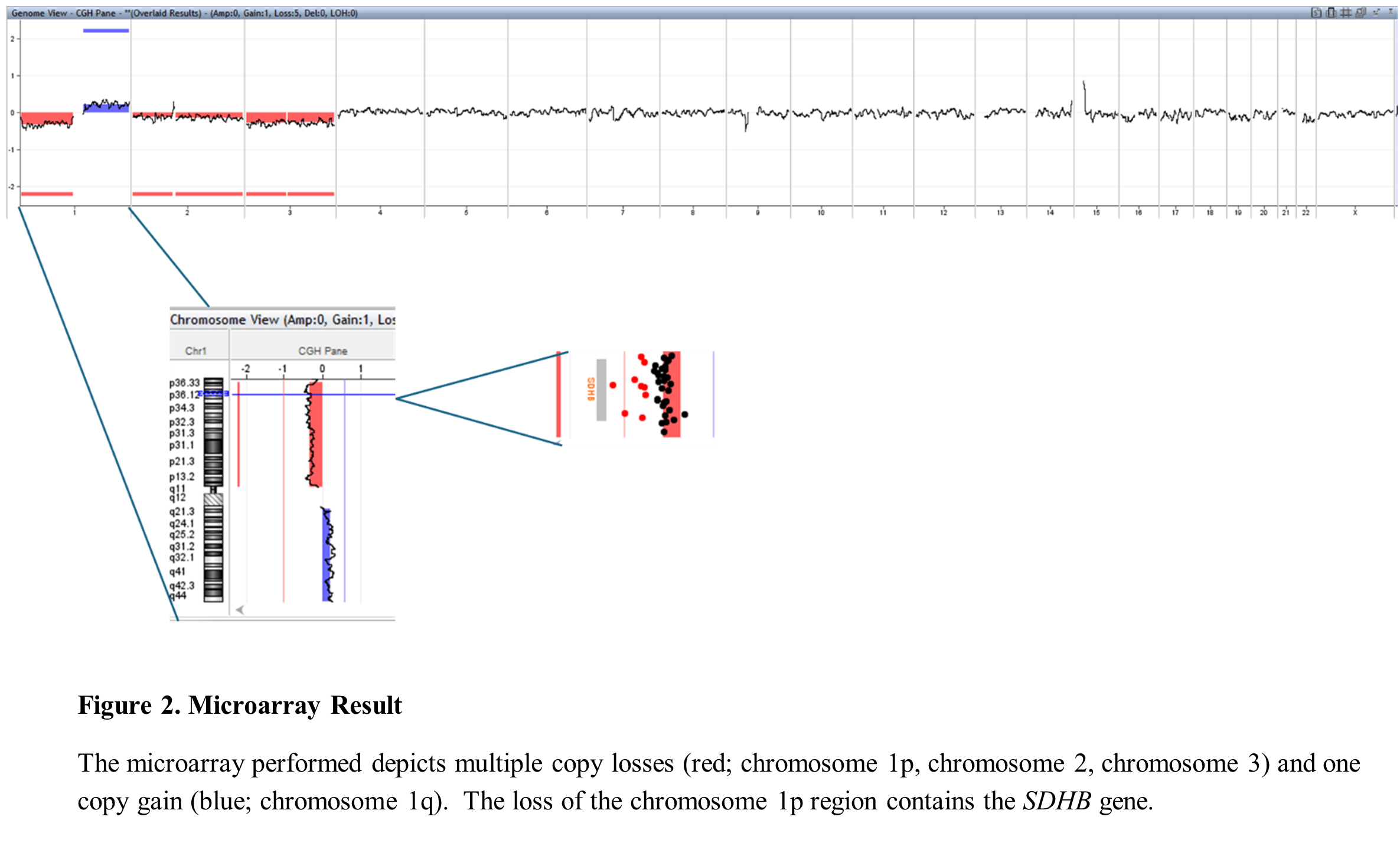
In-house microarray analysis of DNA isolated from tumor was performed (Figure 2). Multiple losses and gains were observed including a loss of the entire p-arm of chromosome 1, encompassing the SDHB gene in the 1p36.13 region. A complete loss of 1p in all cells would exhibit a log ratio of -1, whereas in this case, a log ratio of -0.3 suggested the presence of loss in ~30% of cells. Because this loss is mosaic, and because loss of an entire arm of chromosome 1 would have been incompatible with life, this chromosome arm loss – and resulting SDHB deletion – were classified as somatic.
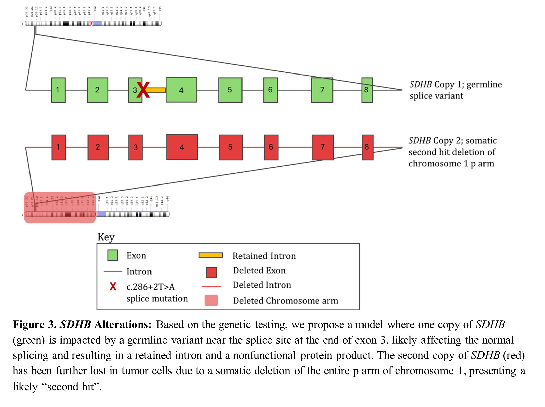
Given the age at presentation, the size of the largest tumor, and the presence of potential metastases or multiple primary lesions, germline testing was pursued. Next generation sequencing (NGS) was performed at an outside facility as part of germline comparator testing. The NGS results were “positive for germline cancer-associated sequence variation in SDHB”. The variant, reported as NM_003000.3 (SDHB): c.286+2T>A p.? was predicted to disrupt the normal splicing of the SDHB gene and result in loss of normal protein function due to the nonsense-mediated decay. This variant has been previously reported in multiple individuals with paraganglioma (Jochmanova et al., 2017). The presence of this variant in both tumor (56%) and non-tumor tissue (46%) confirmed its germline origin. NGS analysis also showed the “somatic” copy number loss of SDHB gene, corresponding to the 1p loss detected by the initial microarray.
Case Resolution
The suprarenal mass was resected and signed out as “SDHB-related paraganglioma”. Angiolymphatic invasion was present. Given that there was no direct involvement of the lesion with the adrenal gland, it was classified as a paraganglioma rather than a pheochromocytoma. Based on the imaging and prior biopsy results, there are likely lesions of the liver, bone, and possibly lung. While liver and lung have rare, reported cases of primary paraganglioma (Liao et al., 2018; Warren et al., 2022), bone does not contain paraganglia. Thus, lesions in bone are likely to represent true metastasis. Given the presence of angiolymphatic invasion on the largest tumor resection in addition to the size and distribution of lesions, it is still plausible that the liver and lung lesions are metastases from the largest paraganglioma. Clinically, metastases and multiple primaries will be managed in the same manner, so this distinction is of a more academic interest. Interestingly, following diagnosis, it was found that several of the patient’s paternal family members have had pheochromocytoma or paraganglioma with known SDHB pathogenic variant.
Discussion
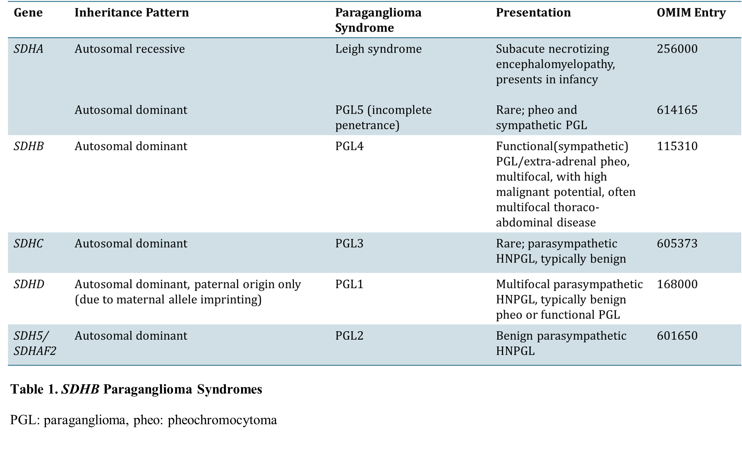
Paragangliomas (called pheochromocytoma when arising from the adrenal medulla) are neoplasms of the paraganglia. They can be sympathetic or parasympathetic, functional or nonfunctional, and can occur in both pediatric and adult patients. Previously believed to be mostly sporadic, it is now estimated that approximately 35% of paragangliomas are related to germline pathogenic variants (Raygada et al., 2011). These include variants in the succinate dehydrogenase (SDH) family of genes. The four known genes in the family, SDHA, SDHB, SDHC, and SDHD have each been linked to a distinct paraganglioma-related syndrome (summarized in Table 1) (Baysal et al., 2015; Kantorovich et al., 2010).
Amar et al previously suggested that germline genetic testing for pheochromocytoma and functional paraganglioma be guided by the clinical phenotype. If there is a known family history or syndromic presentation, testing should begin with the suspected gene (e.g. RET, VHL, SDHD). If the tumor is apparently sporadic but with certain unusual features, testing should follow a logical sequence based on known genotype-phenotype associations. Specifically mentioned are VHL testing for bilateral tumors and SDHB testing for tumors with extra-paraganglionic metastases. (Amar et al., 2005) Because SDH pathogenic variants may be present in up to 30% of apparently sporadic cases, SDHB, SDHC, and SDHD immunohistochemistry can be used as a preliminary test to suggest further genetic testing of the tumor and the patient (van Nederveen et al., 2009).
In this patient, genetic testing was initiated before family history was revealed. While this initially appeared to be a sporadic tumor, the presence of several atypical characteristics (young age, large tumor size, and multiple extra-adrenal lesions) suggested a genetic predisposition. Clinical suspicion was high for either a pheochromocytoma or a functional sympathetic paraganglioma (based on urine metanephrines) with metastases or multiple primary thoraco-abdominal paragangliomas. Because of these atypical characteristics and markedly decreased SDHB immunohistochemical staining pattern, SDHB and SDHD testing were suggested.
Initial genetic testing of the tumor by microarray found multiple somatic copy number alterations including loss of one copy of SDHB (Figure 2). It would be tempting to attribute the immunohistochemical loss of SDHB staining and the tumorigenesis entirely to this event. However, because of the unusual phenotype of the tumor, it was important to continue searching for a germline predisposing variant as the next step. Comparing the tumor genome either to an adjacent normal tissue or to another patient specimen (blood, saliva, or skin biopsy) can determine if any variants are present in the patient’s entire body (germline) or only in the tumor (somatic). In this patient, the disease can be explained by a model where initial germline splicing variant in the SDHB gene inactivated one of the two functional copies of the gene in all cells. This left the remaining copy of the gene vulnerable to a second hit, which occurred with the deletion of the p-arm of chromosome 1 (Figure 3). This case serves as an example of the importance of considering germline predisposition in a scenario of high suspicion, even if a somatic mutation has already been identified. With advances in genomic techniques, somatic and germline variants can be determined by comparative testing of tumor and non-tumor tissues as performed in this patient.
References
Amar, L., Bertherat, J., Baudin, E., Ajzenberg, C., Bressac-De Paillerets, B., Chabre, O., Chamontin, B., Delemer, B., Giraud, S., Murat, A., Niccoli-Sire, P., Richard, S., Rohmer, V., Sadoul, J. L., Strompf, L., Schlumberger, M., Bertagna, X., Plouin, P. F., Jeunemaitre, X., & Gimenez-Roqueplo, A. P. (2005). Genetic testing in pheochromocytoma or functional paraganglioma. Journal of Clinical Oncology, 23(34), 8812–8818. https://doi.org/10.1200/JCO.2005.03.1484
Baysal, B. E., Maher, E. R., Baysal, B. E., & Maher, E. R. (2015). 15 YEARS OF PARAGANGLIOMA: Genetics and mechanism of pheochromocytoma–paraganglioma syndromes characterized by germline SDHB and SDHD mutations. Endocrine-Related Cancer, 22(4), T71–T82. https://doi.org/10.1530/ERC-15-0226
Jochmanova, I., Wolf, K. I., King, K. S., Nambuba, J., Wesley, R., Martucci, V., Raygada, M., Adams, K. T., Prodanov, T., Fojo, A. T., Lazurova, I., & Pacak, K. (2017). SDHB-related pheochromocytoma and paraganglioma penetrance and genotype–phenotype correlations. Journal of Cancer Research and Clinical Oncology 2017 143:8, 143(8), 1421–1435. https://doi.org/10.1007/s00432-017-2397-3
Kantorovich, V., King, K. S., & Pacak, K. (2010). SDH-related pheochromocytoma and paraganglioma. Best Practice & Research Clinical Endocrinology & Metabolism, 24(3), 415–424. https://doi.org/10.1016/j.beem.2010.04.001
Liao, W., Ding, Z. Y., Zhang, Binhao, Chen, L., Li, G. X., Wu, J. J., Zhang, Bixiang, Chen, X. P., & Zhu, P. (2018). Primary functioning hepatic paraganglioma mimicking hepatocellular carcinoma: A case report and literature review. Medicine, 97(17), e0293. https://doi.org/10.1097/MD.0000000000010293
Mete, O., Asa, S. L., Gill, A. J., Kimura, N., de Krijger, R. R., & Tischler, A. (2022). Overview of the 2022 WHO Classification of Paragangliomas and Pheochromocytomas. Endocrine Pathology, 33(1), 90–114. https://doi.org/10.1007/s12022-022-09704-6
Raygada, M., Pasini, B., & Stratakis, C. A. (2011). Hereditary Paragangliomas. Advances in Oto-Rhino-Laryngology, 70, 99. https://doi.org/10.1159/000322484
Unger, P., Hoffman, K., Pertsemlidis, D., Thung, S., Wolfe, D., & Kaneko, M. (1991). S100 protein-positive sustentacular cells in malignant and locally aggressive adrenal pheochromocytomas. Archives of Pathology & Laboratory Medicine, 115(5), 484–487. https://pubmed.ncbi.nlm.nih.gov/1673596/
van Nederveen, F. H., Gaal, J., Favier, J., Korpershoek, E., Oldenburg, R. A., de Bruyn, E. M., Sleddens, H. F., Derkx, P., Rivière, J., Dannenberg, H., Petri, B. J., Komminoth, P., Pacak, K., Hop, W. C., Pollard, P. J., Mannelli, M., Bayley, J. P., Perren, A., Niemann, S., … Dinjens, W. N. (2009). An immunohistochemical procedure to detect patients with paraganglioma and phaeochromocytoma with germline SDHB, SDHC, or SDHD gene mutations: a retrospective and prospective analysis. The Lancet. Oncology, 10(8), 764. https://doi.org/10.1016/S1470-2045(09)70164-0
Warren, B. C., Bond, T., Hessler, R., & Boldt, J. (2022). Primary Paraganglioma of the Lung in an Asymptomatic Patient. Cureus, 14(6). https://doi.org/10.7759/cureus.25562