Case 1172: A Man in His Fourth Decade with a Thyroid Nodule
Authors: André Lametti and Simion Chiosea
Date of Publication: April 1, 2026
Clinical History
A man in the fourth decade of life with unremarkable past medical and family history presents to medical attention for abdominal pain and is found to be hypercalcemic. Subsequent investigations reveal a serum parathyroid hormone (PTH) level over three times the upper limit of normal.
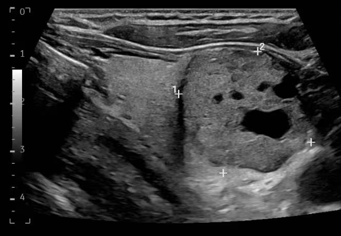
Ultrasound of the neck shows a thyroid nodule (Figure 1) which is aspirated and classified cytologically as atypia of undetermined significance. Molecular testing of the aspirate is positive for expression of PTH messenger RNA, suggesting parathyroid origin.
Figure 1: Ultrasound image of the neck demonstrates an approximately 3 cm heterogeneous nodule in the thyroid.
The patient agrees to thyroid lobectomy for the removal of a presumed intrathyroidal parathyroid gland. Surgery is uncomplicated and after the simultaneous removal of the affected thyroid lobe and an ipsilateral enlarged parathyroid gland, intraoperative serum PTH levels drop by approximately 90%.
Gross Description
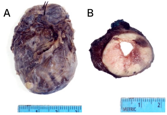
The specimen consists of a slightly enlarged thyroid lobe weighing 20 grams and containing a single well-demarcated tan nodule measuring 2.6 cm in greatest dimension. There is focal hemorrhage and cystic change (Figure 2). The background parenchyma appears normal.
Figure 2: Gross image of the thyroid lobe and isthmus. A. Outer aspect and thyroid "capsule" before sectioning. B. Cut section of the nodule demonstrates discoloration and heterogeneity but clear demarcation from thyroid parenchyma.
The separately excised parathyroid is enlarged but otherwise grossly unremarkable.
Microscopic Examination
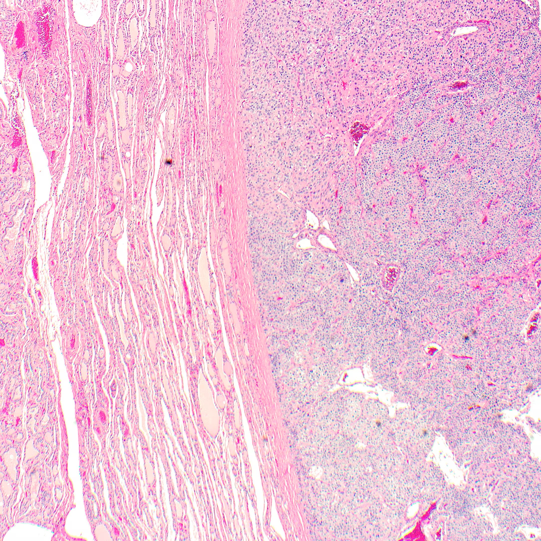
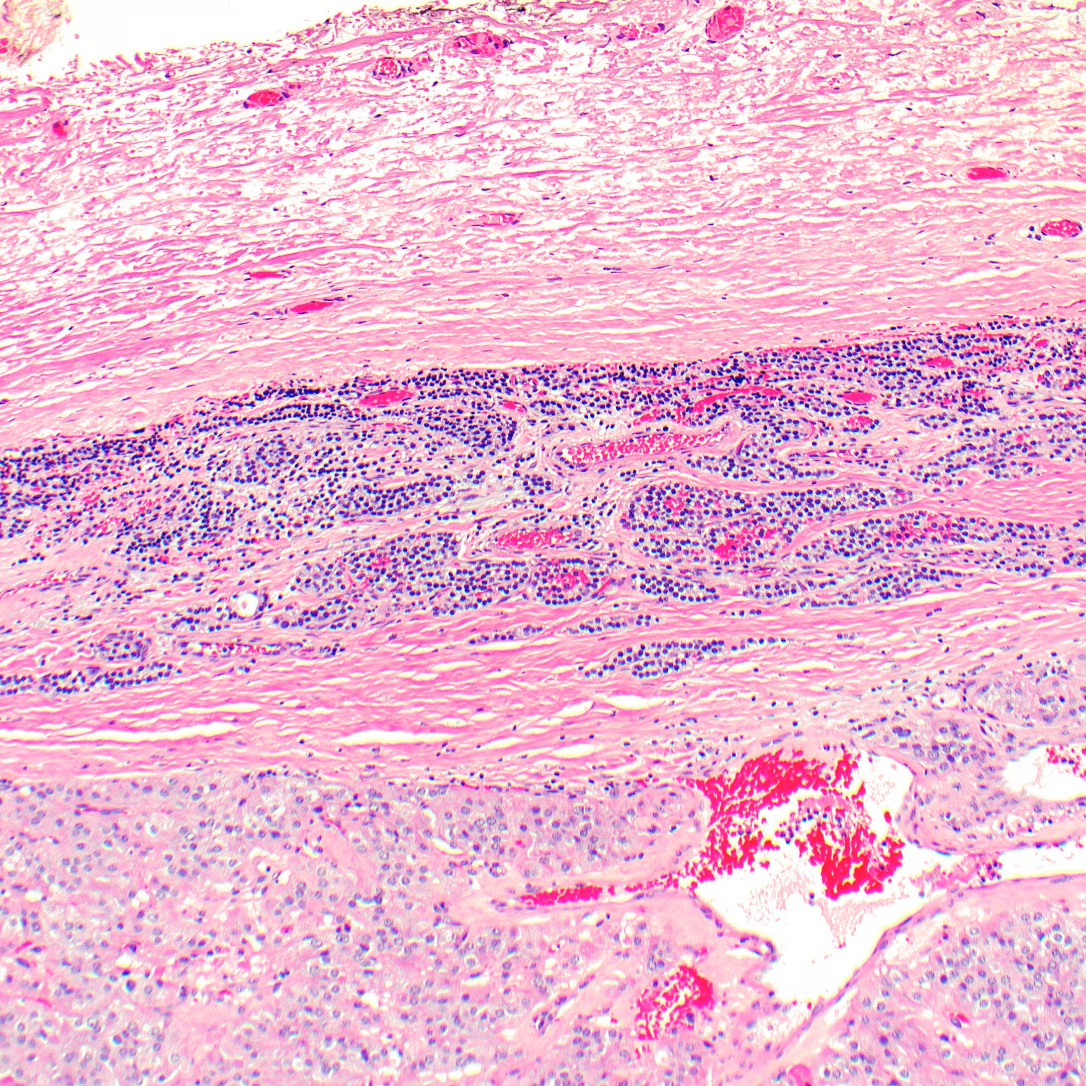
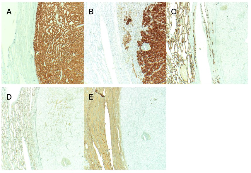
The nodule is encapsulated and clearly distinct from the background thyroid (Figure 3). Along the part of the capsule that is not in contact with thyroid parenchyma, a focus of normal-appearing parathyroid tissue is present (Figure 4). The immunoprofile of the nodule is supportive of parathyroid lineage (Figure 5).
Figure 3. Tumor-normal interface. The tumor (right) is separated from the background thyroid (left) by a moderately thick and well-defined capsule (hematoxylin and eosin, 40× original magnification).Figure 4. Background parathyroid. Normal-appearing parathyroid tissue (center) is present between the encapsulated tumor (bottom) and soft tissue of the neck (top; hematoxylin and eosin, 100× original magnification).Figure 5. Immunohistochemical lineage markers to distinguish thyroid and parathyroid origin. PTH and chromogranin A are positive in the nodule (right of each photomicrograph), while PAX8, TTF1 and thyroglobulin are positive in the background thyroid (left of each photomicrograph) but not in the nodule. A. PTH, 40× original magnification. B. Chromogranin A, 40× original magnification. C. PAX8, 40× original magnification. D. TTF1, 40× original magnification. E. Thyroglobulin, 40× original magnification.
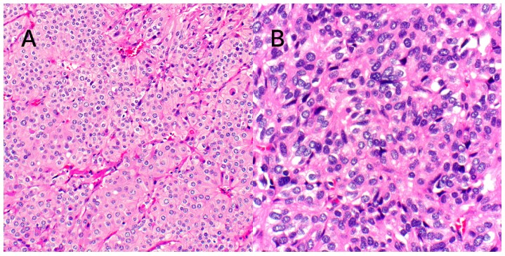
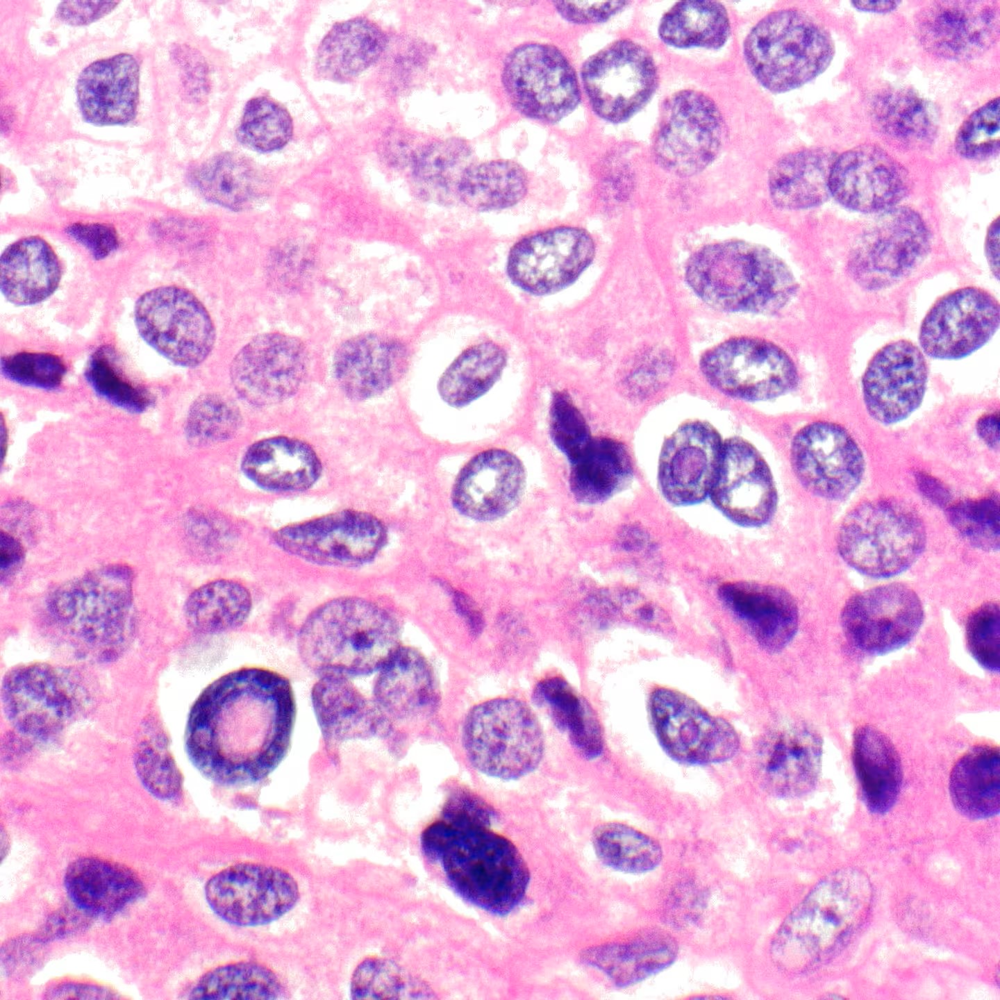
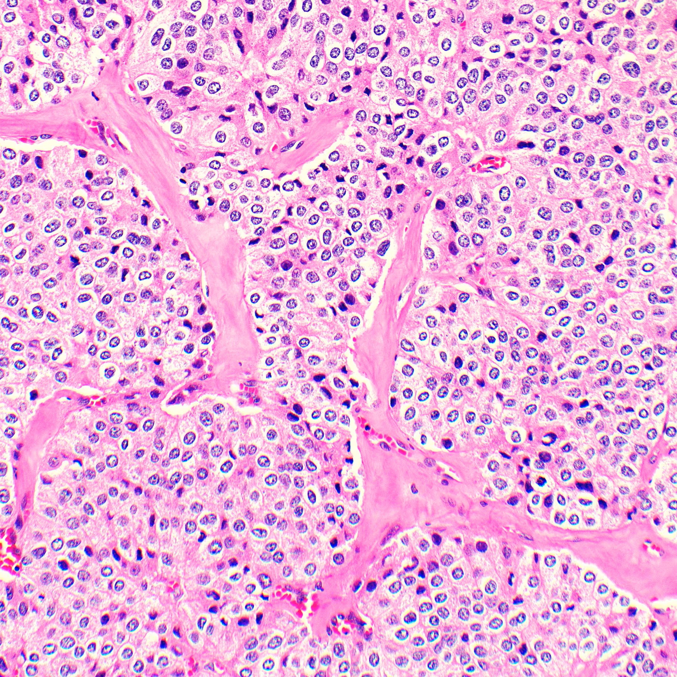
The majority of the tumor is composed of oncocytoid cells with enlarged and mildly atypical nuclei (Figure 6-A). In some areas there is increased nuclear atypia, less prominent oncocytic cytology and increased nuclear-cytoplasmic ratio (Figure 6-B). Intranuclear pseudoinclusions are relatively easy to identify (Figure 7).
Figure 6. Cytomorphology of the tumor cells. A. Most of the tumor consists of oncocytoid cells with marginated chromatin and mild nuclear size variation (hematoxylin and eosin, 200× original magnification). B. Certain areas display oblong cells with more enlarged nuclei and coarse chromatin (hematoxylin and eosin, 400× original magnification).Figure 7. Intranuclear pseudoinclusions. These are easily identified in this case. Pseudoinclusions are not unique to papillary thyroid carcinoma and are seen in a wide spectrum of neoplasia, both endocrine and non-endocrine (hematoxylin and eosin, 600× original magnification).
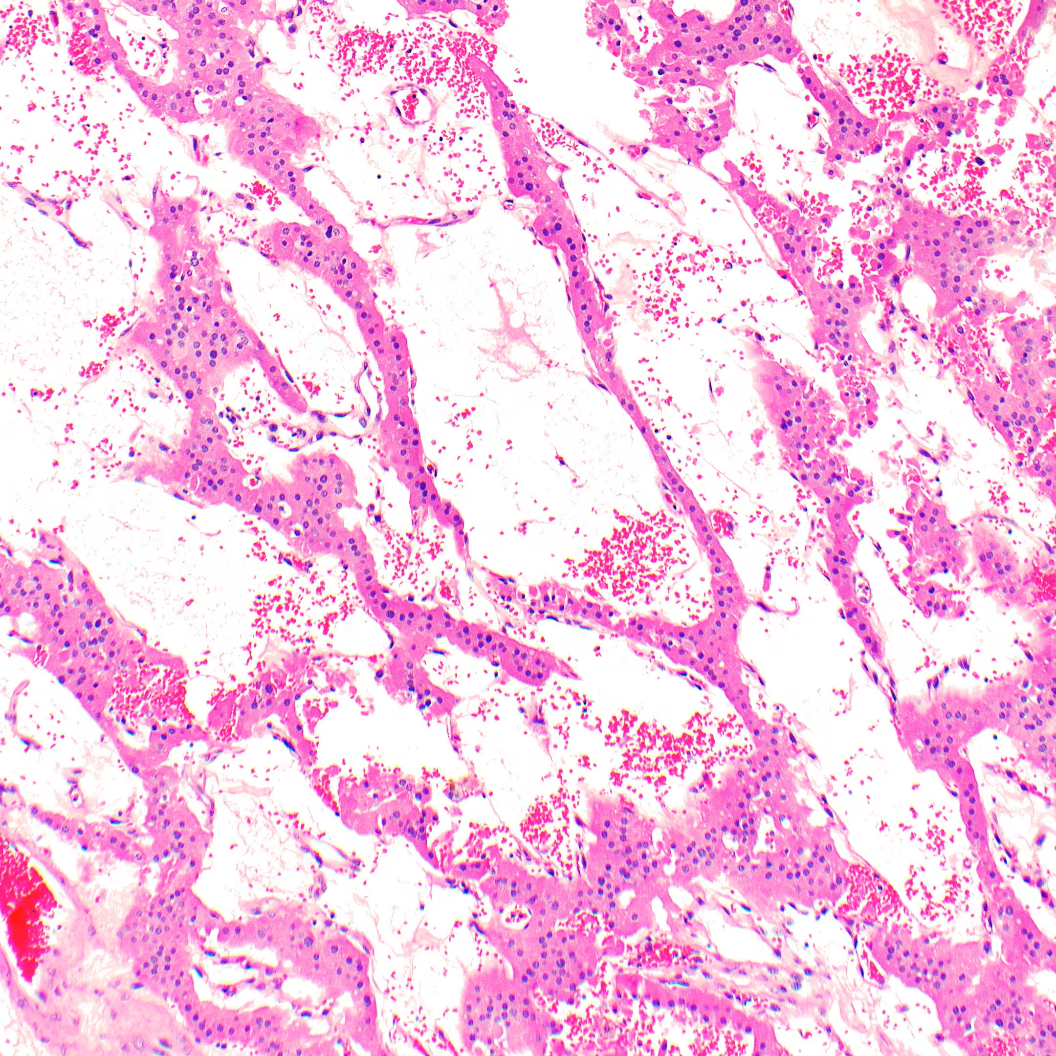
A few areas exhibit cystic degeneration or interstitial edema, likely explaining the gross heterogeneous appearance (Figure 8). There is incompletely developed intratumoral fibrosis (Figure 9).
Figure 8. Edema. Areas with interstitial fluid and blood, the former removed by processing, explain grossly softer areas (hematoxylin and eosin, 100× original magnification).Figure 9. Intratumoral fibrosis. When widespread and especially band-like (not seen here), fibrosis is an atypical feature in parathyroid neoplasia (hematoxylin and eosin, 200× original magnification).
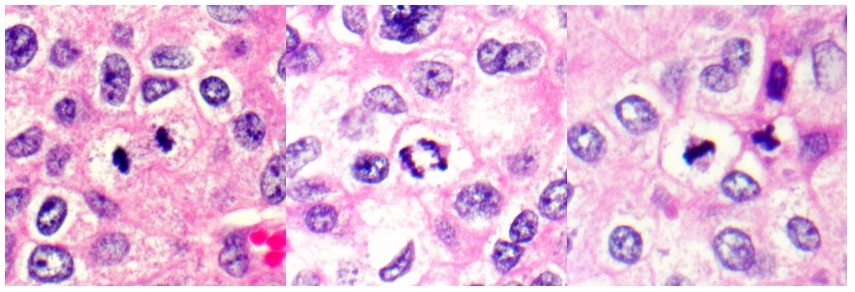
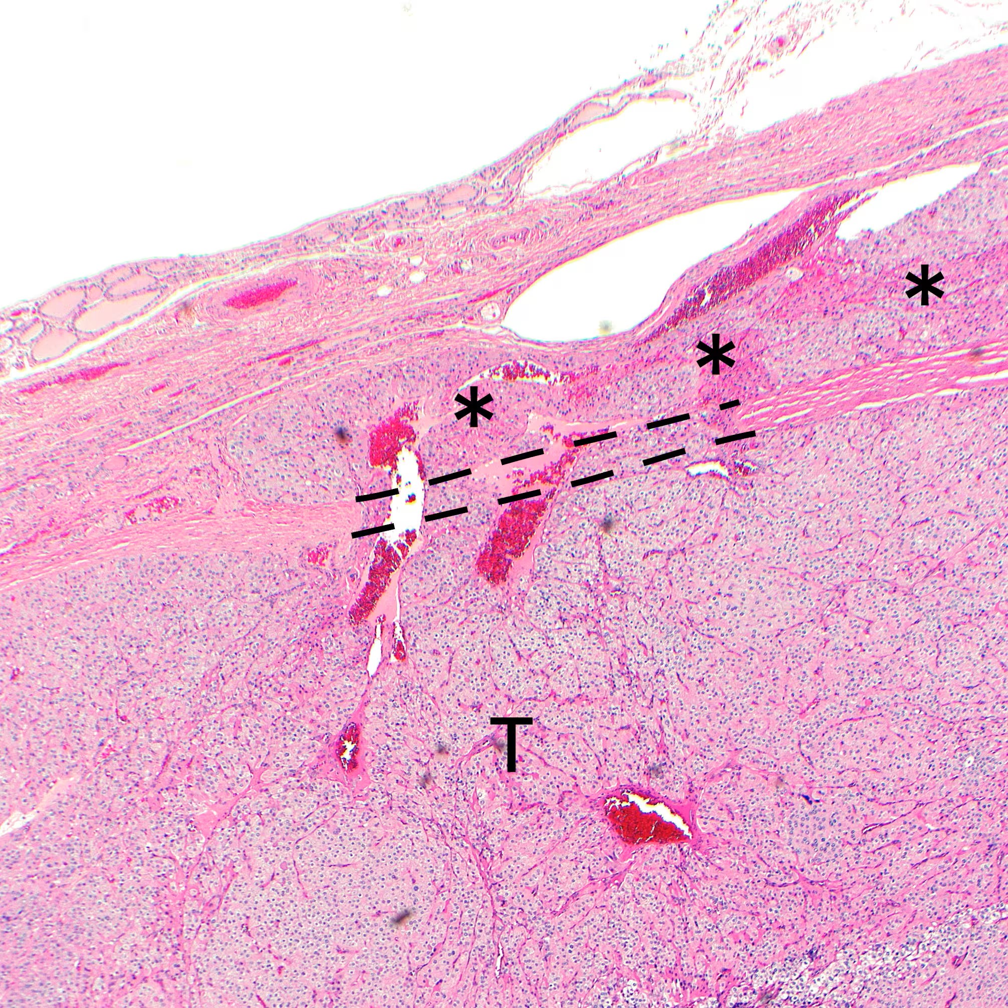
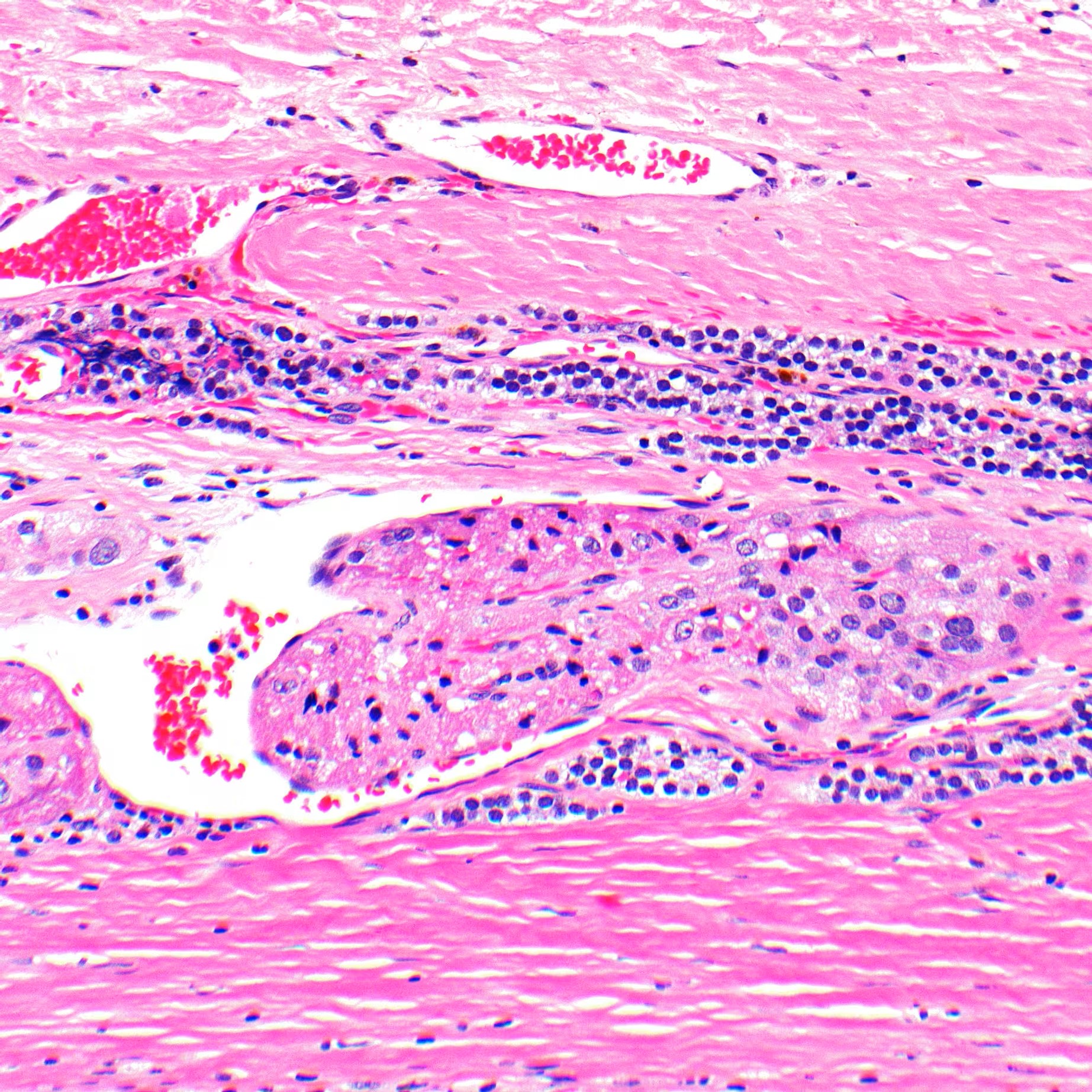
Mitotic activity is readily appreciable, but there are no atypical forms (Figure 10). There is one focus of capsular invasion (Figure 11) and one focus of extratumoral vascular invasion with thrombus formation and endothelialization (Figure 12). There are no additional tumor nodules nor is there infiltration into the thyroid parenchyma. Unequivocal tumor necrosis is not seen.
Figure 10. Mitotic activity. There is increased mitotic activity, but all forms are typical (hematoxylin and eosin, 600× original magnification).Figure 11. Capsular invasion. Tumor (T) creates a breach (dashed lines) in the original tumor capsule and fungates through it (✱) without frank invasion of surrounding structures (hematoxylin and eosin, 20× original magnification).Figure 12. Vascular invasion. Tumor cells are present in a vessel outside of the nodule, with associated fibrin and endothelialization (hematoxylin and eosin, 200× original magnification).
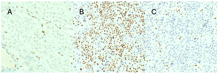
The tumor cells demonstrate variable loss of RB1 and 5-hydroxymethylcytosine (5hmC; Figure 13). p53 and parafibromin stains are also performed but are not definitively interpretable. The Ki-67 proliferation index by image analysis is just under 2%.
Figure 13. Abnormal loss of protein expression. There is variable loss of RB1 and 5hmC (a surrogate for TERT alteration). Endothelial cells are positive in all three photomicrographs and serve as an internal control of normal expression. A. Area with loss of RB1, 200× original magnification. B. Area with retained 5hmC, 200× original magnification. C. Area with loss of 5hmC, 200× original magnification.
The separately removed parathyroid is enlarged and hypercellular but does not show any atypical features.