
Authors: Udit Naik, M.D. and Jon Davison, M.D.
Date of Publication: May 1, 2026
Clinical History
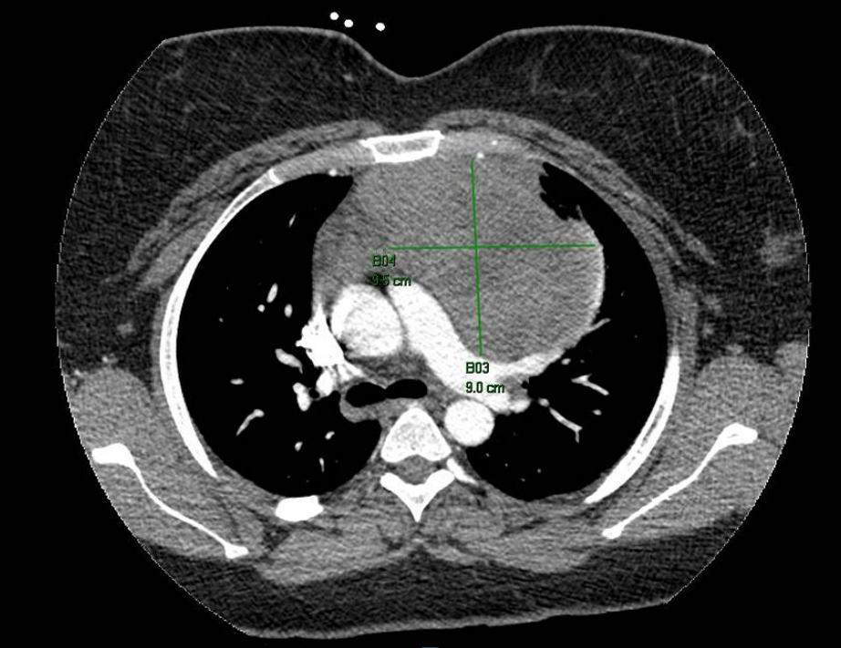
Woman in her 20s presented with an intermittent chest pain which exacerbated on inspiration and a newly developed dry cough. On CT angiogram for chest, she was found to have a 9.5 x 9.0 x 8.0 cm mass arising in anterior mediastinum. CT guided core needle biopsy was performed.
Results
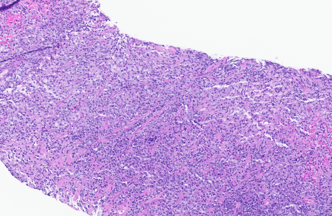
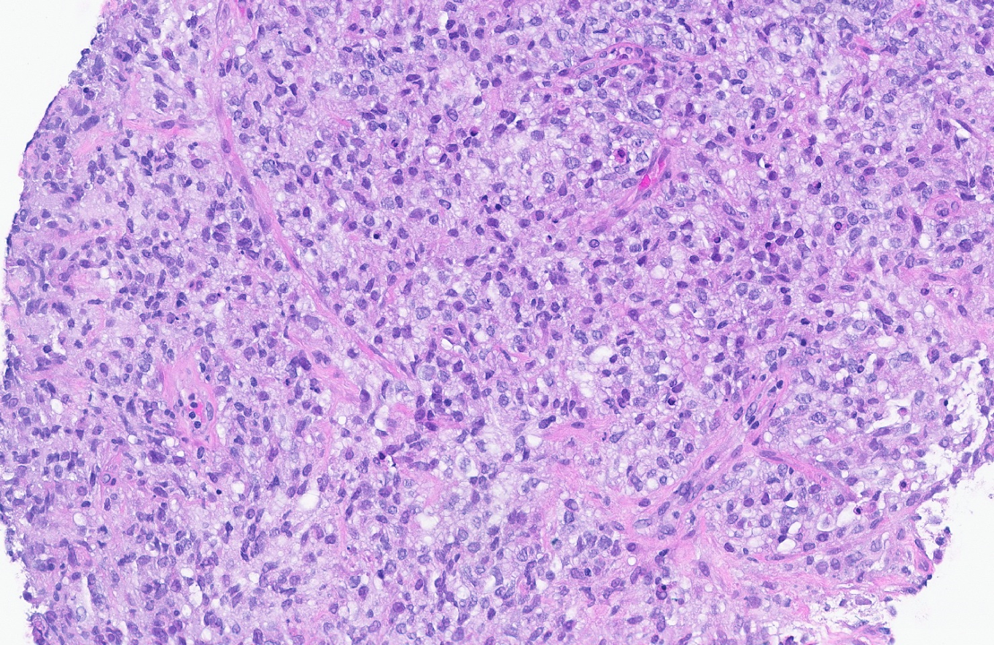
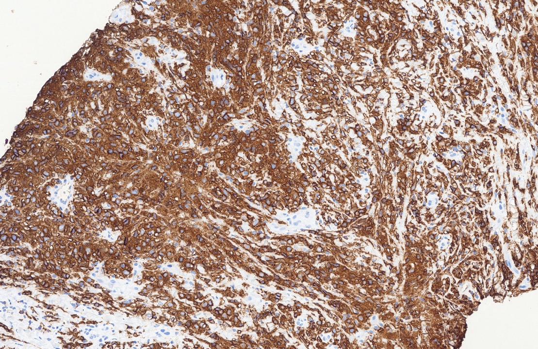
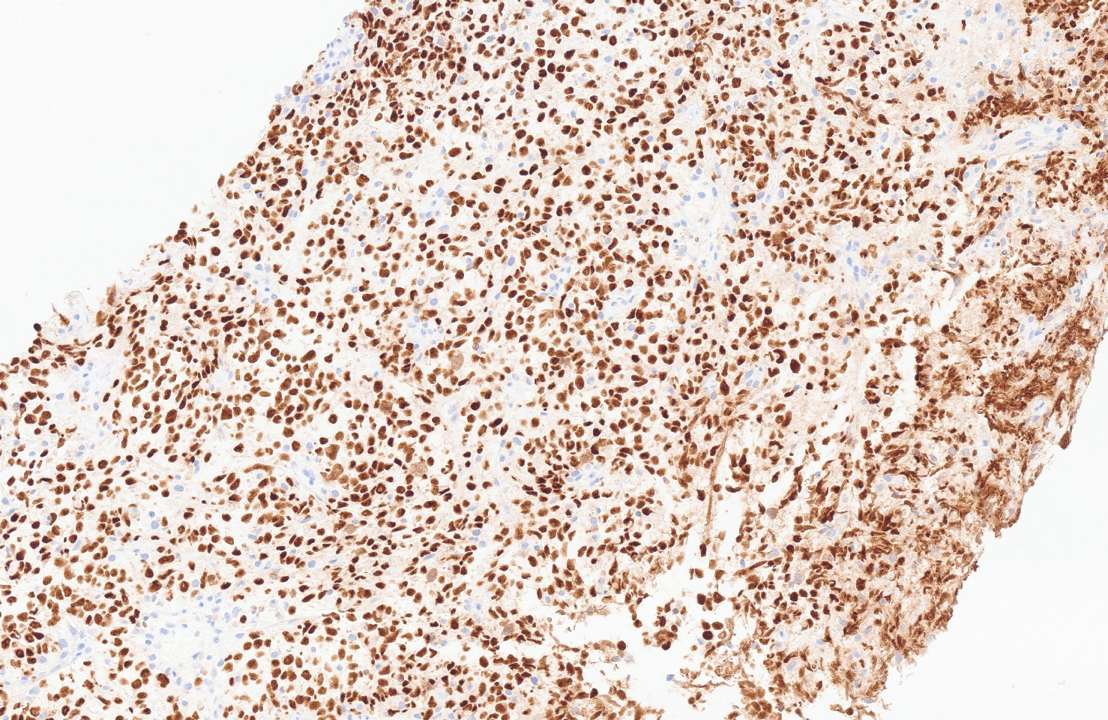
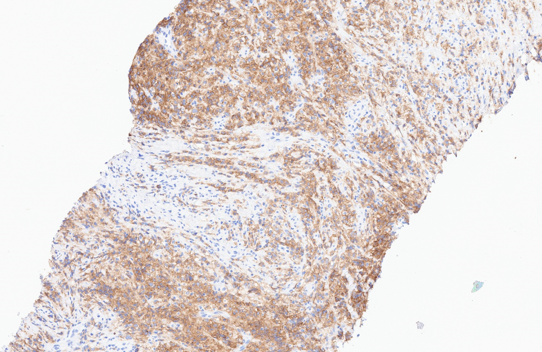
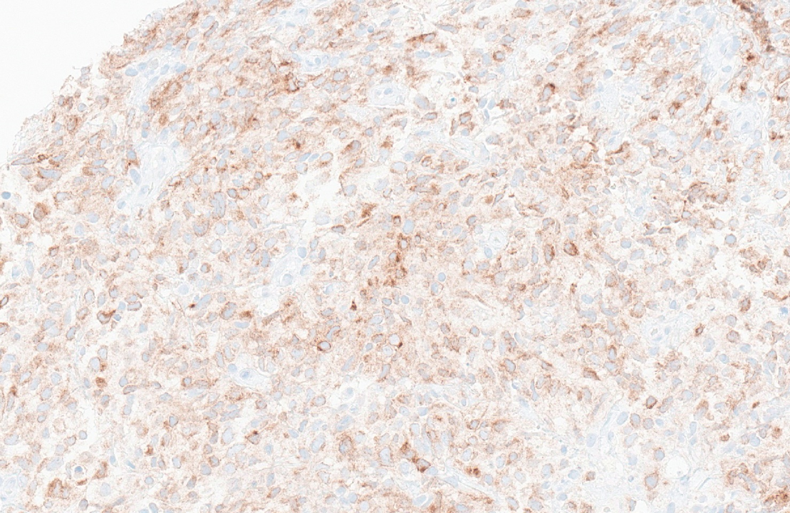
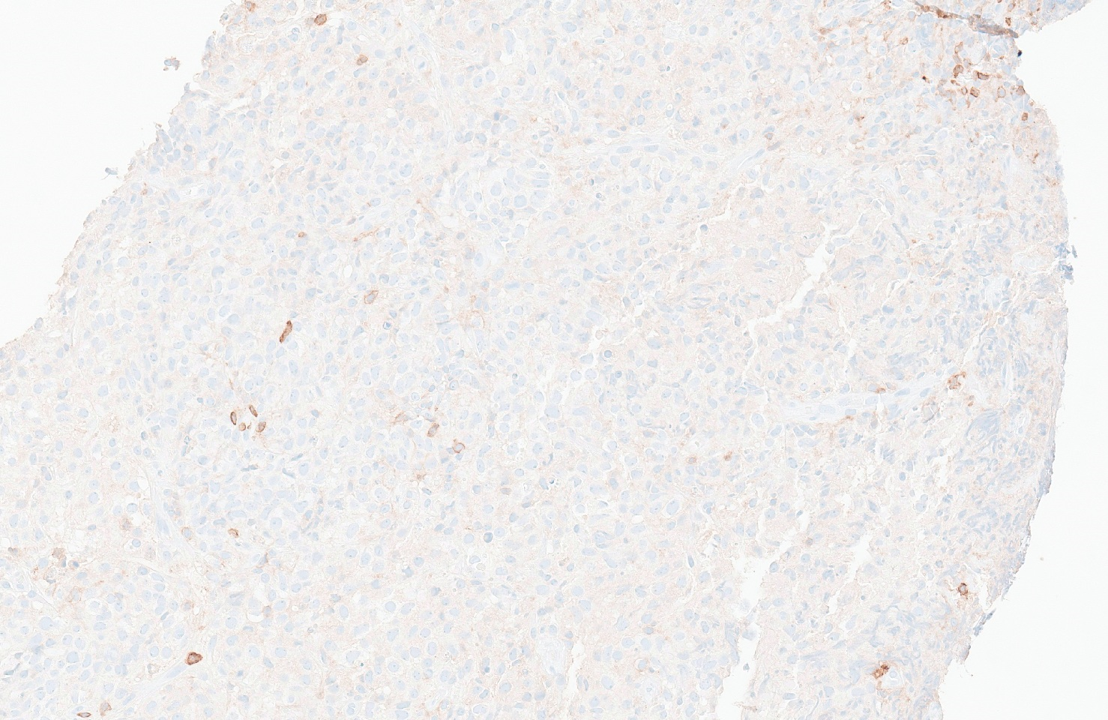
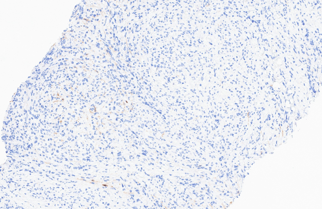
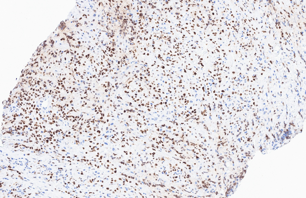
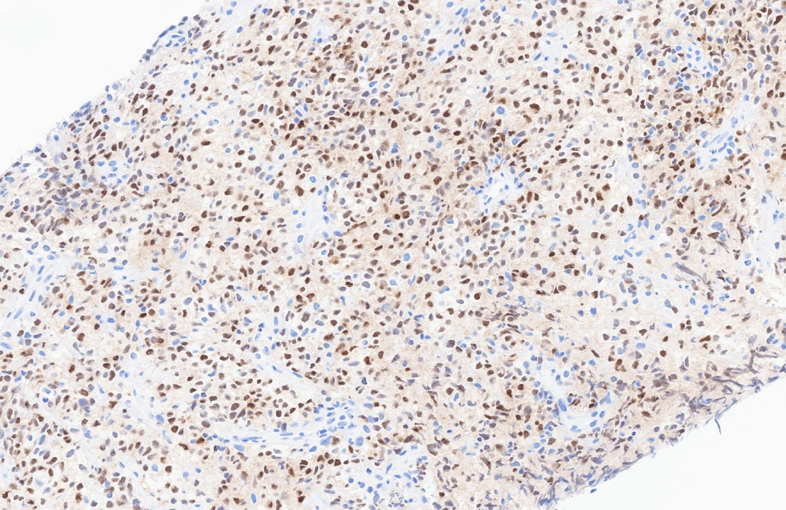
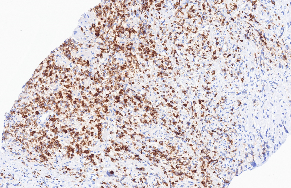
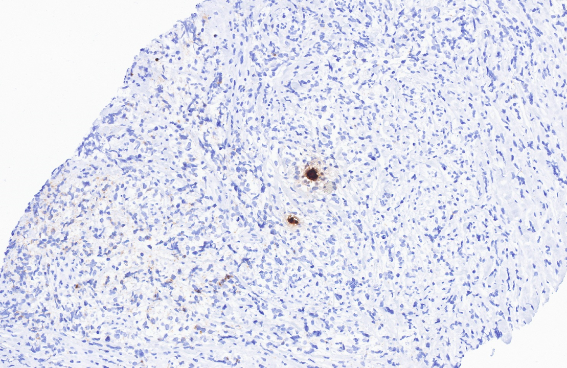
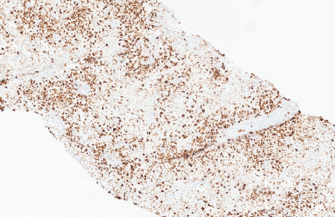
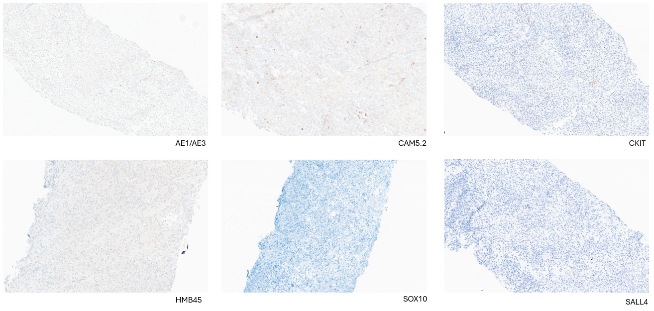
Morphological assessment showed diffuse infiltrate with large cells with moderate amount of clear cytoplasm in a vague organoid pattern with surrounding sclerosis. The tumor cells were strongly positive for CD20, CD19, PAX5, BCL6, BCL2 and CD23, whereas weakly positive for MUM1 and CD30 (variable). The cells are negative for CD10, cyclin-D1, CD5, HHV8 and EBER. The cells express kappa ISH and are negative for lambda ISH. Ki67 highlighted moderate number of cells. Extensive work-up to rule out other differential diagnosis showed that the tumor cells were negative for AE1/AE3, CAM 5.2, OCT 3/4, SALL4, GATA-3, INSM1, synaptophysin, C-kit, SOX-10, HMB-45, Melan-A, desmin, CD34, CD45, CD68, CD163, S-100, LCA, CD99, ALK, CD1a, CD2, CD4, CD21, CD138, TdT, MPO, lysozyme, caldesmon, MyoD1, NUT, Myogenin, NKX2.2 and ERG. The cells retained INI1 and BRG1 expressions. Molecular and cytogenetic investigations were performed. Mutations in EZH2, B2M, SOCS1, STAT6, KMT2D, PALB2, FAT1 genes were identified.
Discussion
Large B-cells with non-germinal center type phenotype (positive for MUM1, negative for CD10) harboring aberrancies such as CD23, CD30 expression and mutations in genes STAT6 and SOCS1 were identified in the mediastinal mass. These findings strongly favored a diagnosis of primary mediastinal large B-cell lymphoma (PMBCL). Presence of B-cell antigens (CD20, C19. CD79a, PAX5) and transcription factors (BOB1, OCT2) and variable expression of CD30 help differentiate this entity from grey zone lymphoma and classic Hodgkin lymphoma. DLBCL-NOS does not usually express CD30, CD23 and can be excluded. Extensive workup helped rule out other commonly differentials - thymoma/thymic carcinoma, teratoma and unusual entities such as melanoma, Ewing sarcoma, NUT carcinoma, SWI/SNF complex deficient undifferentiated carcinoma, Langerhans cell histiocytosis, Rosai-Dorfman disease, among others.
Conclusion
PMBCL presents as a diffuse infiltration of large lymphoid cells, often in a fibrotic background in a young population. Commonality of ‘round blue cell’ morphology in mediastinum in vast variety of entities demands extensive workup. It is critical to differentiate PMBCL from grey zone lymphoma, classic Hodgkin lymphoma and DLBCL due to therapeutic and prognostic implications.