Authors: Mohamed M. Elmoryah, MD and Azfar Neyaz, MD
Date of Publication: July 1, 2026
Clinical History
An adult male in his thirties with no significant past medical history presented with a rapidly developing forearm mass. The lesion had been present for approximately 1 to 2 weeks and was noted to fluctuate in size. The patient denied pain, trauma, erythema, sensory deficits, weakness, systemic symptoms, or additional masses. Physical examination demonstrated a mobile, spherical area of induration on the radial aspect of the forearm. There was no overlying erythema, warmth, or neurovascular compromise.
Radiologic Findings
Ultrasound showed a hypoechoic lobulated subcutaneous mass measuring approximately 1.9 cm with adjacent vascularity. MRI demonstrated a well-defined 1.8 cm subcutaneous mass within the volar aspect of the distal forearm. The lesion was considered nonspecific, with differential considerations including a myxoid soft tissue neoplasm or vascular lesion.
Histologic Findings
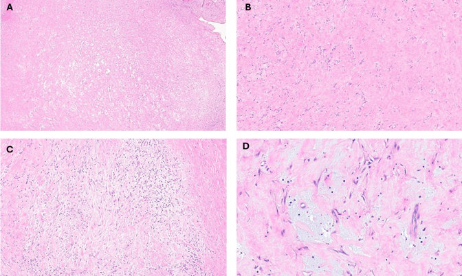
Microscopic examination shows a poorly circumscribed, variably cellular soft tissue lesion with a myxoid to fibrous appearance on low-power examination. The lesion is composed of loosely arranged spindle cells set within a myxoid stroma, demonstrating a characteristic “tissue culture–like” growth pattern, along with scattered inflammatory cells and collagen bundles. On high-power examination, the tumor cells are plump spindle to stellate shaped with vesicular nuclei and indistinct cytoplasm embedded in a myxocollagenous matrix, focally accompanied by extravasated erythrocytes and inflammatory cells. No significant cytologic atypia, necrosis or mitotic activity is present (Figure 1 A-D).
Ancillary Studies
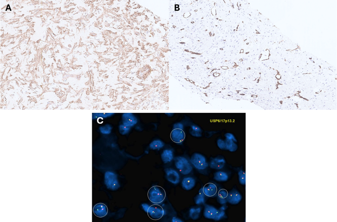
Immunohistochemical studies show that the lesional cells are positive for SMA, consistent with myofibroblastic differentiation, while desmin, S100, cytokeratins, and CD34 are negative (Figure 2A & 2B). FISH analysis demonstrates a USP6 gene rearrangement (Figure 2C), confirming the diagnosis.