
Authors: Jerome Givi, MD; Gabriela M. Quiroga-Garza, MD
Date of Publication: October 1, 2026
Clinical History
An adult male in his sixties with significant cardiopulmonary and hepatic comorbidities was found to have a left renal mass concerning for renal cell carcinoma. The lesion had been identified on prior imaging, although follow-up was not established. Subsequent imaging demonstrated interval enlargement of the renal mass with development of retrocrural and retroperitoneal lymphadenopathy concerning for metastatic disease. A percutaneous biopsy of the renal mass was performed.
Radiologic Findings
Initial MRI demonstrated a 2.9 cm heterogeneously T2-hypointense, enhancing lesion involving the anterior lower pole of the left kidney without macroscopic fat or evidence of local invasion. Subsequent imaging initially showed relative stability, followed by interval enlargement to approximately 4.1 cm. The later study also demonstrated poor opacification of the left renal vein and infrarenal inferior vena cava with venous expansion and multiple enlarged retrocrural and retroperitoneal lymph nodes concerning for metastatic disease. An additional indeterminate small lesion was present in the contralateral kidney.
Histologic Findings
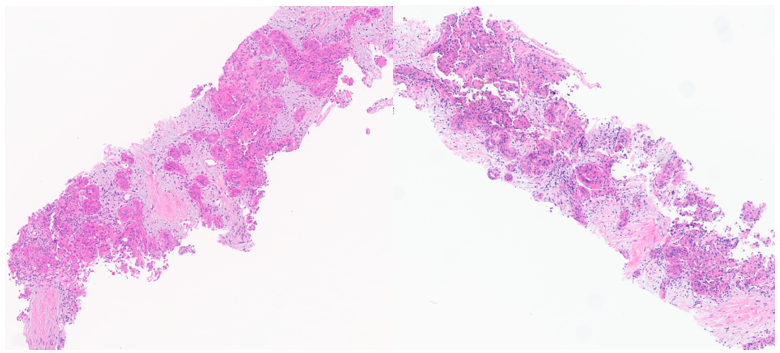
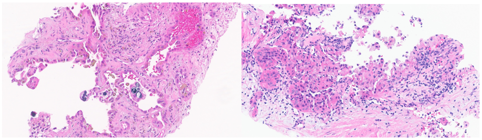
Microscopic examination of the renal mass biopsy shows a high-grade carcinoma composed predominantly of large polygonal cells with abundant eosinophilic cytoplasm, enlarged nuclei, and conspicuous nucleoli. The neoplastic cells are arranged in small nests and vaguely papillary structures separated by variably dense fibrous stroma. Focal cystic areas and psammomatous calcifications are also present (Figure 1A-D).
Ancillary Studies
Immunohistochemical studies demonstrate expression of AE1/AE3, CK7, alpha-methylacyl-CoA racemase (AMACR), and PAX8, supporting renal epithelial differentiation. The neoplastic cells are negative for CA9 and c-kit, while colloidal iron is non-contributory.
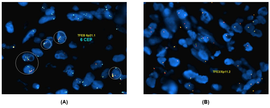
Results of FISH studies are shown in Figure 3.
Diagnosis and Discussion