
Authors: Emma S. Harvey, DO; Octavia M. Peck Palmer, PhD, FADLM
Date of Publication: October 1, 2026
Introduction
Waldenström macroglobulimenia (WM), a relatively rare subtype of lymphoplasmacytic lymphoma comprised of IgM-producing B-cells, has a known association with cryoglobulinemia (1). Diagnostic laboratory evaluation of WM includes quantitative immunoglobulin levels, serum protein electrophoresis, and serum immunofixation, among other tests; however, these tests rely on the immunoglobulins being unbound in the serum, meaning that cryoprecipitation can lead to false negative results, depending on how cool the blood sample becomes. In this case report, we review a common pitfall in laboratory diagnostics for patients with WM: the presence of a cryoglobulin.
Clinical History
A patient in their 8th decade of life with no significant past medical or surgical history presented to their primary care physician for a 10-month history of syncopal episodes with no known triggers, associated visual changes, or palpitations. These syncopal episodes precipitated multiple falls and mechanical injury, including an ankle fracture which prompted them to seek medical care. In addition to syncopal episodes, the patient noted dizziness, weakness, and lightheadedness. Laboratory findings at the time were notable for leukocytosis (WBC 2.6 k/uL, reference range: 3.8k - 10.8 k/uL). The patient was referred to cardiology and orthostatic vitals, an EKG, an echocardiogram, and ambulatory cardiac monitoring were ordered. Summarily, findings were consistent with mild chronic hypotension and hyperdynamic left ventricular function, and midodrine was initiated.
Later that month, the patient was admitted to the hospital for their third syncopal episode. Their blood was noted to be viscous during blood draw. A CBC revealed a new, mild anemia (Hgb 10.5 g/dL, reference range: 11.6-14.6 g/dL), and a peripheral smear showed thrombocytosis, with a platelet count of 600-750 k/uL (reference range: 150-450 k/uL). They were discharged on aspirin therapy with a referral to Hematology/Oncology, with intentions to trend their platelet count. At the Hematology/Oncology follow-up appointment, a repeat platelet count was 513k. Additional history revealed fatigue and B symptoms; at this time, peripheral blood flow cytometry was ordered. Results were suggestive of a low-level B-cell lymphoproliferative disorder, and a myeloid NGS panel showed an ASX L1 mutation. A bone marrow biopsy showed a low level of involvement by an IgM+ plasmacytic population. During this outpatient workup for hematologic malignancy, the patient presented to the emergency department three times due to non-resolving, spontaneous epistaxis.
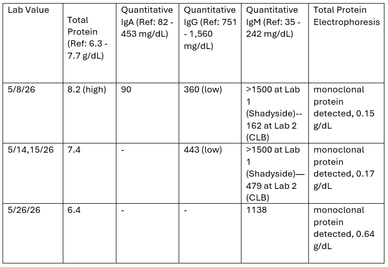
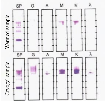
Due to findings consistent with plasma cell neoplasm, serum protein electrophoresis (SPEP), serum immunofixation electrophoresis (IEP), quantitative IgA, quantitative IgG, and quantitative IgM were ordered. Immunoglobulin quantitation was run at the same hospital as collection (Shadyside laboratory, Lab 1) and showed an IgM level that was over the detectable limit (>1500.0 mg/dL, reference range: 35 - 242 mg/dL). IgG was low at 360 mg/dL (reference range: 751 - 1,560 mg/dL), and IgA was within normal limits at 9.0 mg/dL (reference range: 82 - 453 mg/dL). The sample was sent to the medical center’s central lab (Clinical Laboratory Building, Lab 2) for repeat IgM testing, which was now low at 162 mg/dL. The SPEP, performed at Lab 2, did show a monoclonal protein at 0.15 g/dL; IEP showed this to be type IgM kappa. Due to these discrepant results, another sample was drawn a week later; again, the IgM level was unable to be quantified at Lab 1, since it was over the detectable limit. Sending the sample to Lab 2 showed elevated IgM at 479 mg/dL (Table 1).
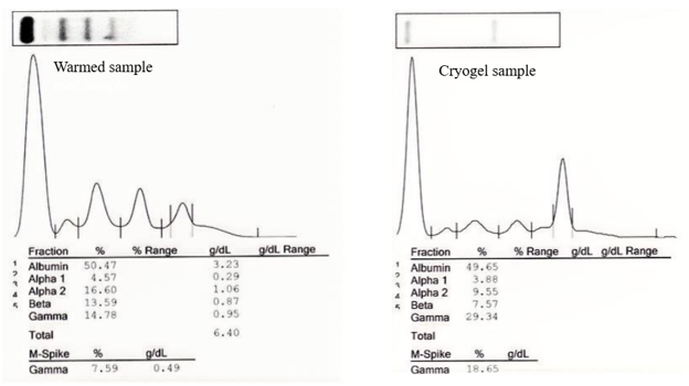
At this time, a peripheral smear was reviewed, which showed agglutination vs. Rouleaux formation. Notably, the hematopathologist mentioned that cryoglobulin-like material could not be excluded. A polyspecific antihuman globulin test, cold hemagglutinins, cryoglobulin screen with reflex to cryoglobulin type, and a hepatitis B and C test were ordered. The antihuman globulin test, cold hemagglutinin test, and hepatitis B and C tests were all negative, though the cryoglobulin screen was positive, and subsequent cryoglobulin typing showed Type II cryoglobulinemia with monoclonal IgM kappa and polyclonal IgG (44.07 mg/dL IgG, 7.0 mg/dL IgA, 294 mg/dL IgM). Two weeks later, an additional sample was sent to Lab 2 for IgM quantitation, along with TPE (Fig. 1) and IEP (Fig. 2). Now, the sample was warmed and showed IgM levels of 1138 mg/dL.
Discussion
Cryoglobulins are defined as immunoglobulins that precipitate at temperatures less than 37◦C, but dissolve upon warming. Due to their temperature lability, laboratory handling is of the utmost importance for proper detection. In this case, when the sample was processed at the same institution where it was drawn, the sample presumably did not cool appreciably from the patient’s body temperature. Thus, IgM and IgG were detectable. However, because the quantitative IgM value was critically high, it was sent to another institution for confirmational testing. Transportation of the sample allowed for cooling of the blood and precipitation of antibody complexes, precluding appropriate quantitation of IgM. After suspicion was raised for the presence of cryoglobulins, the specimen was warmed prior to IgM and IgG quantitation, revealing an immunoglobulin fraction that had previously been undetectable due to formation of immune complexes.
To test for cryoglobulins, avoiding premature cryoprecipitation is key. The blood sample must be kept at 37◦C during transport to the laboratory. The sample is then allowed to clot and is centrifuged for serum separation before cooling. If present, the cryoglobulins will precipitate, and can be extracted, rinsed, and characterized and quantified by immunofixation and Western blotting (2).
Cryoglobulinemia, especially type II cryoglobulinemia, is not always symptomatic (3); thus, clinical suspicion for the presence of cryoglobulins may be low. Luckily, in our case, the sample was tested soon enough after collection to detect hypergammaglobulinemia, and the discrepant results following repeat testing prompted further consideration. However, in this case, the ramifications of a falsely low IgM could be twofold, as it would have delayed the diagnosis of our patient’s WM and cryoglobulinemia.
Tables and Figures
References
- Hobbs M, Fonder A, Hwa YL. Waldenström Macroglobulinemia: Clinical Presentation, Diagnosis, and Management. J Adv Pract Oncol. 2020;11(4):381-389. doi:10.6004/jadpro.2020.11.4.5
- Roccatello D., Saadoun D., Ramos-Casals M. et al. Cryoglobulinaemia. Nat Rev Dis Primers 4, 11 (2018). https://doi.org/10.1038/s41572-018-0009-4
- Desbois A. C., Cacoub P., Saadoun D. Cryoglobulinemia: An update in 2019. Joint Bone Spine, Volume 86, Issue 6, 2019, Pages 707-713, ISSN 1297-319X, https://doi.org/10.1016/j.jbspin.2019.01.016.