
Authors: Zachary High, MD and Samer Khader, MD
Date of Publication: November 1, 2026
Case Summary
A man in his 50s with a history of parathyroid carcinoma with invasion into the thyroid gland (status post right thyroid lobectomy) underwent routine biannual ultrasound surveillance of his neck. Multiple enlarged lymph nodes within the right cervical level 2 nodal chain were discovered, the largest measuring 4.3 cm. No discrete abnormalities were observed in the right thyroid bed or isthmus. The left thyroid lobe was notable for one 0.4 cm cystic nodule with benign features.
His cervical lymphadenopathy was palpable on physical examination. The patient was otherwise asymptomatic, reporting no dyspnea, dysphagia, voice changes, fevers, night sweats, or unintended weight loss. His most recent calcium and parathyroid hormone levels were within normal limits.
An ultrasound-guided lymph node biopsy was performed.
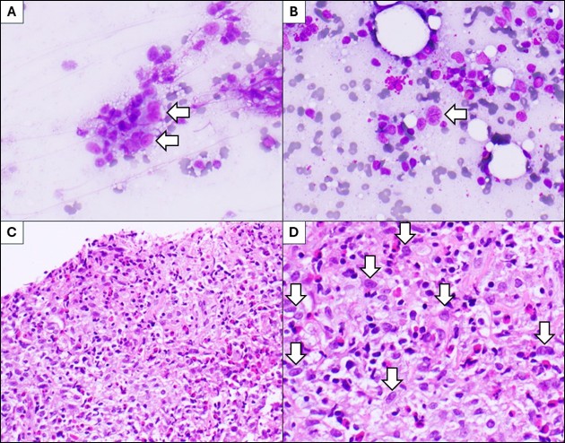
Microscopic examination of the Diff-Quik stained direct smears and H&E cell block sections scattered large atypical mononuclear and binuclear cells with a moderate to abundant amount of cytoplasm and enlarged irregular nuclei. The atypical cells were present within a mixed inflammatory infiltrate composed of small lymphocytes, eosinophils, plasma cells, and histiocytes (Figure 1).
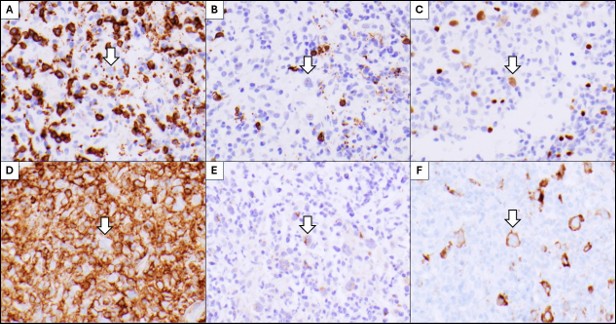
The enlarged mononuclear and binuclear cells stained positive for CD15 and CD30 (membranous with accentuation in the Golgi area of the cytoplasm), weak positive nuclear for PAX5, and negative for CD3, CD20, and CD45 (Figure 2). The atypical cells were additionally negative for BOB1, OCT2, EBER, PTH, and AE1/3.