
Final Diagnosis
Intentional Ethylene Glycol Exposure
Discussion
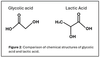 This patient presented with findings characteristic of ethylene glycol toxicity including altered mental status, anion gap metabolic acidosis with an osmolal gap, and hypocalcemia. A slight elevation in lactic acid is expected in the setting of ethylene glycol toxicity as it is metabolized into glycolaldehyde and glycolic acid by alcohol dehydrogenase and aldehyde dehydrogenase, respectively, to produce pyruvate which is further converted to lactic acid. Interestingly, our patient had discrepant lactic acid measurements with one method (whole blood lactate) measuring over 4 fold higher concentration as compared to the other (i-Stat). This falsely elevated lactic acid is secondary to interference from glycolic acid, one of the toxic metabolites of ethylene glycol. This elevation is recognized in the literature as a “lactate gap” and clinically interpreted as evidence supporting ethylene glycol ingestion (1-3). This interference occurs in laboratory systems which detect lactic acid using a lactate oxidase system. This system measures hydrogen peroxide produced when lactic acid is converted to pyruvate by lactate oxidase. Due to structural similarities, glycolic acid can also be used by lactate oxidase to generate hydrogen peroxide and thus be interpreted as lactic acid (Figure 2). An alternative measurement system using lactate dehydrogenase is not susceptible to this form of interference (1-3).
Acid-base homeostasis is critical for normal physiology but despite complex regulation and buffering systems, derangements and imbalances in the acid-base equilibrium occur. Initial evaluation of an acid/base imbalance begins by analyzing the direction of the pH change. A pH below 7.35 indicates acidemia whereas a pH above 7.45 indicates alkalemia (4-5). These disturbances can result from various metabolic and respiratory processes, which can be evaluated based on the CO2 and bicarbonate concentrations. In an acidotic patient, an elevated CO2 suggests a respiratory cause whereas a low level suggests a metabolic cause for the acidosis (4-5). Additionally in metabolic acidosis, the bicarbonate concentration is decreased reflecting the increase in hydrogen ion plasma concentration. A relative increase in hydrogen plasma ion concentration as compared to base can occur in the setting of increased endogenous acid production, exogenous acid ingestion, decreased acid excretion, or increased renal or GI bicarbonate losses (4-5). Calculating an anion gap and osmolar gap can further narrow the cause of metabolic acidosis.
Plasma neutrality necessitates a balance of cations and anions. If there is an increase in measured anions as compared to cations this leads to an anion gap. Characteristic causes of an increased anion gap metabolic acidosis include methanol, uremia, diabetic ketoacidosis, propylene glycol, paracetamol, paraldehyde, isoniazid, lactic acidosis, and ethylene glycol (4-5). Another helpful parameter in determining the cause of a metabolic acidosis is osmolality. Serum osmolality is determined by the concentration of dissolved solutes in the plasma and can be calculated using the sodium, urea, glucose, and ethyl alcohol concentrations (4-5). It can also be determined in the laboratory by a freezing point depression. A difference in the calculated and measured osmolality defines an osmolal gap and indicates there are solutes present in the plasma that are not accounted for in the typical osmolality equation (4-5).
In our case the patient presented with a low pH indicating there was an ongoing acidosis which was determined to be metabolic based on the low bicarbonate and low CO2. A increased anion gap of 24 mmol/L was present along with an osmolar gap. While this pattern could also be seen with methanol ingestion, the volatile alcohol screen was negative (2). Additionally, as discussed above the spuriously elevated lactic acid secondly to glycolic acid interference and low calcium due to the production of oxalic acid with subsequent binding to calcium supported the clinical suspicion for ethylene glycol ingestion (6-7). This was confirmed through the ethylene glycol screen and elevated concentration measurement by gas chromatography. Of note, if there is high clinical suspicion for ethylene glycol ingestion, multiplying the osmolar gap by the molecular weight of ethylene glycol can given an estimated total ethylene glycol concentration. In our case this resulted in an estimated concentration of 347.2 mg/dL as compared to the actual final concentration of 332 mg/dL.
Ethylene glycol is a sweet tasting but odorless compound which as a parent molecule is relatively non-toxic but will result in an osmolal gap early after ingestion (6-7). It is subsequent metabolism and production of toxic metabolites, glycolic and oxalic acid which lead to the clinical symptoms and characteristic anion gap acidosis. Ethylene glycol belongs to the group of toxic alcohols and is found in many easily accessible household products (2, 6-7). It is responsible for accidental exposures, primarily among children, and intentional exposures/overdoses in the setting of suicide or as a substitute for ethanol among individuals with chronic alcohol use disorder (6-7).
 The clinical presentation of ethylene glycol ingestion is variable based on the amount, timing, and co-ingestion of other substances. The typical progression of clinical symptoms includes neurologic manifestations often starting within 30 minutes of ingestion which can include signs of intoxication, altered mental status, somnolence, coma, and seizures (2,6-7). Cardiopulmonary complications manifest around 12-24 hours with renal complications secondary to calcium oxalate crystal deposition including flank pain, oliguria/anuria, gross hematuria, and renal failure occurring 24-72 hours after ingestion (2,6-7).
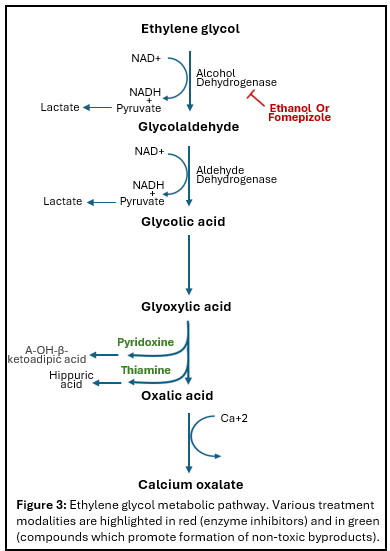
Treatment for ethylene glycol ingestion is multipronged with the overarching goal of correcting the underlying acidosis and preventing additional formation of toxic metabolites (Figure 3). Ethanol and more recently fomepizole can be used to competitively inhibit alcohol dehydrogenase and prevent downstream metabolism of ethylene glycol (2,6-7). While there is less evidence supporting their efficacy, thiamine and pyridoxine are given to shunt ethylene glycol metabolism toward the non-toxic metabolites, alpha oh beta ketoadipic acid and hippuric acid, respectively (2,6-7) . Lastly in the setting of severe acidosis with ethylene glycol levels greater than 50 mg/dL, severe electrolyte derangements, or renal dysfunction, dialysis can be initiated. Bicarbonate can also be given for severe acidosis and calcium repleted for symptomatic hypocalcemia including seizures, tetany, QT prolongation (2 & 7). In our case, the patient was initially given bicarbonate to correct the acidosis and started on fomepizole, thiamine, and pyridoxine while waiting for dialysis. These resulted in an appropriate decrease in ethylene glycol and improvement in mental status.
References
1. Sagar AS, Jimenez CA, Mckelvy BJ. Lactate gap as a tool in identifying ethylene glycol poisoning. BMJ Case Rep 2018;2018(doi:10.1136/bcr-2018-224243
2. Kraut JA, Mullins ME. Toxic Alcohols. N Engl J Med 2018;378(3):270-280, doi:10.1056/NEJMra1615295
3. Kamboj M, Bejjanki H, Gopal S, et al. Erroneously High Lactate: A Guide to Diagnosing Ethylene Glycol Poisoning. Biomed Hub 2019;4(2):1-3, doi:10.1159/000499967
4. Burger MK, Schaller DJ. Metabolic Acidosis. [Updated 2023 Jul 17]. In: StatPearls. Treasure Island (FL): StatPearls Publishing; 2024 Jan. Available from: https://www.ncbi.nlm.nih.gov/books/NBK482146/
5. Kraut JA, Madias NE. Metabolic acidosis: Pathophysiology, diagnosis and management. Nat Rev Nephrol 2010;6(5):274-85, doi:10.1038/nrneph.2010.33
6. Porter WH. Ethylene glycol poisoning: quintessential clinical toxicology; analytical conundrum. Clin Chim Acta 2012;413(3-4):365-77, doi:10.1016/j.cca.2011.10.034
7. Iqbal A, Glagola JJ, Nappe TM. Ethylene Glycol Toxicity. [Updated 2022 Sep 26]. In: StatPearls. Treasure Island (FL): StatPearls Publishing; 2024 Jan. Available from: https://www.ncbi.nlm.nih.gov/books/NBK537009/
8. Rastogi A, Itagaki B, Bajwa M, et al. Spurious elevation in serum creatinine caused by ingestion of nitromethane: implication for the diagnosis and treatment of methanol intoxication. Am J Kidney Dis 2008;52(1):181-7, doi:10.1053/j.ajkd.2007.12.010
9. Beal S, Nazarullah A, Sanford K. Quick Compendium of Clinical Pathology. In: (Mais D. ed.) American Society for Clinical Pathology: Canada; 2018; pp. 1-54.