
Final Diagnosis
Zellweger Spectrum Disorder (ZSD) [OMIM #214100]
Discussion
Genetic testing revealed heterozygosity in PEX1 for a sequence variant c.2528G>A, predicted to result in amino acid substitution p.Gly843Asp. a common pathogenic variant. The patient was also found to be heterozygous in PEX1 for a sequence variant c.3655C>T, predicted to result in premature protein termination (p.Gln1219*), which has not been reported in the literature, but is likely pathogenic. The genetic testing combined with the clinical and biochemical findings confirmed the diagnosis of Zellweger Spectrum Disorder (ZSD).
Prevalence
The incidence of ZSD may vary by region. Measures in the United States estimate an incidence of 1:50,0001,2,3, however more recent data obtained from a New York State newborn screening laboratory utilizing a biochemical assay (CP26:0-LPC) detected an incidence of 1:133,000 in the population studied1. Other observed regional variations include an incidence of 1:12,191 live births in a French-Canadian region of Quebec and an incidence of 1:500,000 births in Japan1,3,4.
Pathogenesis
Peroxisomes are single membrane bound organelles with near ubiquitous expression in human cells; they are particularly abundant in liver and kidney5. PEX genes encode peroxins required for peroxisomes biogenesis. Genes and corresponding peroxin function are summarized below1,11,12:
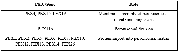
Peroxisomes participate in numerous essential roles including: beta-oxidation of VLCFAs, alpha oxidation of branched chain fatty acids, amino acid and ethanol catabolism, biosynthesis of bile acids, steroids, gluconeogenesis, formation of plasmalogens and degradation of cytotoxic hydrogen peroxide5. Loss of function leading to accumulation of VLCFA, pristanic and phytanic acid, reduced steroid biosynthesis, and decreased neutralization of cytotoxic hydrogen peroxide fuels the pathogenesis resulting in neuronal membrane injury and demyelination, adrenal damage, retinal degeneration, and liver toxicity among the other sequela that is observed1,3,5.
Clinical Features
The clinical findings in Zellweger syndrome exist on a broad phenotypic spectrum with manifestations often differing based on the age of presentation. However, overlap can occur. Common findings based on three age groups are summarized below1,3,5,10:
Neonates-Infants:
Hypotonia, reduced spontaneous movements and weak cry. Patients frequently have feeding difficulties and seizures which can present early in life. Physical features include facial dysmorphism (high forehead, large fontanelles, wide sutures, hypoplastic supraorbital ridges, and a broad nasal bridge). System specific abnormalities include ocular (glaucoma, retinopathy, cataracts), nervous (gyral malformations and varying degrees of sensorineural hearing loss), hepatobiliary (hepatomegaly, cholestasis, hepatic dysfunction), renal cysts, and skeletal (chondrodysplasia punctata of patella and other long bones).
Older infants and children:
Developmental delays and failure to thrive with or without hypotonia; intellect can be unaffected. Hepatic dysfunction, adrenal insufficiency, hearing and vison impairment and kidney stones (calcium oxalate). Regression of previously attained milestones can also be observed (leukodystrophy)
Adolescents/Adults:
Developmental delays, adrenal insufficiency, leukodystrophy, peripheral neuropathy and cerebellar ataxia.
Clinically the broad phenotypic spectrum of Zellweger Spectrum Disorder is described in terms of “severe” ZSD and “intermediate/milder” ZSD. Historically severe ZSD was referred to as Zellweger syndrome, while intermediate phenotypes were referred to as neonatal adrenoleukodystrophy (NALD) and milder phenotypes as infantile Refsum disease (IRD)1,10. The new designation serves to better highlight the spectrum of disease severity as opposed to the previously assigned categories10. Severe ZSD presents in the neonatal period with the aforementioned clinical features; death typically occurs within the first year due to respiratory complications or infections1. In contrast the intermediate/milder ZSD present later, often secondary to developmental delays but presentation in the newborn period can occur1.
Diagnosis
Suspicion for Zellweger syndrome is raised through a combination of clinical and laboratory findings. The diagnosis is established through molecular testing identifying biallelic pathogenic variants in one of the 13 PEX genes1. A notable exception is mentioned in the genetics section. Salient findings on screening assays are summarized in the table below1,6:
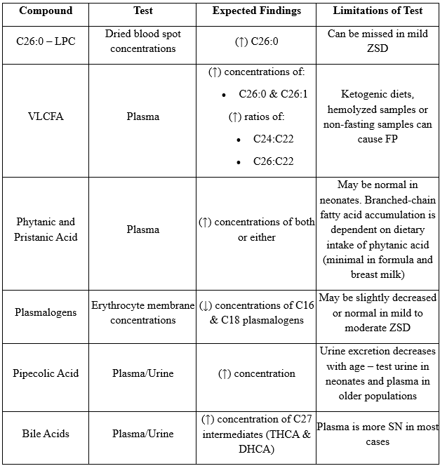
Several options exist for establishing a diagnosis of ZSD using molecular techniques and the choice of testing is often determined by the strength of accompanying biochemical and clinical data1. In scenarios where individuals present with findings suspicious for ZSD, diagnosis is more likely to be obtained through gene targeted testing – where a multigene panel for peroxisome biogenesis disorders is obtained (includes 13 PEX genes)1. In contrast, when findings are not suggestive of a specific diagnosis, identification is often obtained through comprehensive genomic testing – exome sequencing or genome sequencing1.
Genetics
The molecular genetic testing used in Zellweger Spectrum Disorder (ZSD) includes 13 PEX genes1. They include in decreasing order of frequency (percentage of ZSD attributable to pathogenic variants) the following genes: PEX1 (60%), PEX6 (14.5%), PEX12 (7.6%), PEX26 (4.2%), PEX10 (3.4%), PEX2 (3.1%), PEX5 (2.0%), PEX13 (1.5%), PEX16 (1.1%), PEX3 (0.7%), PEX19 (0.6%), PEX14 (0.5%), and PEX11B (0.1%)1,7. Of note, one exception to identifying biallelic pathogenic variants occurs in PEX6 variant p.Arg860Trp; which has been observed with ZSD in a heterozygous state attributable to allelic expression imbalance1.
Genotype-Phenotype Correlations
Homozygosity for PEX1 p.Ile700TyrfsTer42 has been associated with more severe phenotypes1. In contrast, homozygosity for PEX1 p.Gly843Asp has been associated with milder or sometimes intermediate phenotypes with a normal neurologic examination and ocular phenotype reported1,8,9.
Differential Diagnosis
In consideration of other disorders, the differential diagnosis can be broad and will vary based on the age of presentation as well as the prominent symptomatology1,3. Prominent hypotonia in the neonatal period often raises concern for chromosomal abnormalities such as Down Syndrome or Prader-Willi Syndrome, while adrenocortical insufficiency can raise suspicion for X-linked adrenoleukodystrophy3. Other important considerations where overlap in clinical and biochemical findings occur include a group of single peroxisomal enzyme deficiencies including Acyl-CoA oxidase type 1 (ACOX1) and D-bifunctional protein deficiency (DBP); in these cases, a broader peroxisomal multigene panel is recommended for workup1,3.
Management/Treatment
Current management and treatment recommendations for ZSD focus primarily on symptom management and supportive care. Clinical evaluations for the following symptoms are important to perform regularly and include the following: growth failure, deafness, visual impairment, hepatic dysfunction, renal and adrenal insufficiency, and neurological decline3,10. For severe ZSD the primary focus in management includes seizure control with feeding and respiratory support. Additional interventions discussed for less severe cases are also valuable for improved quality of life3,10. Intermediate or milder cases of ZSD also focus on feeding and nutritional support including fat soluble vitamin supplementation, monitoring liver function, hearing and vision deficit support, baseline MRI studies for leukodystrophy monitoring, adrenal insufficiency monitoring with steroid replacement if necessary, and hyperoxaluria monitoring3,10.
Genetic Counseling
Zellweger Spectrum Disorder is inherited in an autosomal recessive manner; the PEX6 variant serving as a notable exception1. After identification of a pathogenic variant in the patient, subsequent molecular testing of the parents is recommended to confirm heterozygosity of a pathogenic PEX variant for reliable risk recurrence assessments1. In detecting carrier status of siblings, molecular workup is recommended as biochemical markers are normal1. Carriers can be offered prenatal or preimplantation genetic testing1,3.
References
[1] Steinberg SJ, Raymond GV, Braverman NE, et al. Zellweger Spectrum Disorder. 2003 Dec 12 [Updated 2020 Oct 29]. In: Adam MP, Feldman J, Mirzaa GM, et al., editors. GeneReviews® [Internet]. Seattle (WA): University of Washington, Seattle; 1993-2024. Available from: https://www.ncbi.nlm.nih.gov/books/NBK1448/
[2] Gould SJ, Raymond GV, Valle D. The peroxisome biogenesis disorders. In: Scriver CR, Beaudet AL, Sly WS, Valle D, eds. The Metabolic and Molecular Bases of Inherited Disease. 8 ed. New York, NY: McGraw-Hill; 2001:3181-218.
[3] Klouwer FC, Berendse K, Ferdinandusse S, Wanders RJ, Engelen M, Poll-The BT. Zellweger spectrum disorders: clinical overview and management approach. Orphanet J Rare Dis. 2015 Dec 1;10:151. doi: 10.1186/s13023-015-0368-9. PMID: 26627182; PMCID: PMC4666198.
[4] Levesque S, Morin C, Guay SP, Villeneuve J, Marquis P, Yik WY, Jiralerspong S, Bouchard L, Steinberg S, Hacia JG, Dewar K, Braverman NE. A founder mutation in the PEX6 gene is responsible for increased incidence of Zellweger syndrome in a French Canadian population. BMC Med Genet. 2012 Aug 15;13:72. doi: 10.1186/1471-2350-13-72. PMID: 22894767; PMCID: PMC3483250.
[5] Elumalai V, Pasrija D. Zellweger Spectrum Disorder. [Updated 2024 Sep 1]. In: StatPearls [Internet]. Treasure Island (FL): StatPearls Publishing; 2024 Jan-. Available from: https://www.ncbi.nlm.nih.gov/books/NBK560676/
[6] Vogel BH, Bradley SE, Adams DJ, D'Aco K, Erbe RW, Fong C, Iglesias A, Kronn D, Levy P, Morrissey M, Orsini J, Parton P, Pellegrino J, Saavedra-Matiz CA, Shur N, Wasserstein M, Raymond GV, Caggana M. Newborn screening for X-linked adrenoleukodystrophy in New York State: diagnostic protocol, surveillance protocol and treatment guidelines. Mol Genet Metab. 2015 Apr;114(4):599-603. doi: 10.1016/j.ymgme.2015.02.002. Epub 2015 Feb 12. PMID: 25724074.
[7] Ebberink MS, Mooijer PA, Gootjes J, Koster J, Wanders RJ, Waterham HR. Genetic classification and mutational spectrum of more than 600 patients with a Zellweger syndrome spectrum disorder. Hum Mutat. 2011;32:59–69.
[8] Poll-The BT, Gootjes J, Duran M, De Klerk JB, Wenniger-Prick LJ, Admiraal RJ, Waterham HR, Wanders RJ, Barth PG. Peroxisome biogenesis disorders with prolonged survival: phenotypic expression in a cohort of 31 patients. Am J Med Genet A. 2004;126A:333–8.
[9] Majewski J, Wang Z, Lopez I, Al Humaid S, Ren H, Racine J, Bazinet A, Mitchel G, Braverman N, Koenekoop RK. A new ocular phenotype associated with an unexpected but known systemic disorder and mutation: novel use of genomic diagnostics and exome sequencing. J Med Genet. 2011;2011;48:593–6.
[10] Braverman NE, Raymond GV, Rizzo WB, Moser AB, Wilkinson ME, Stone EM, Steinberg SJ, Wangler MF, Rush ET, Hacia JG, Bose M. Peroxisome biogenesis disorders in the Zellweger spectrum: An overview of current diagnosis, clinical manifestations, and treatment guidelines. Mol Genet Metab. 2016 Mar;117(3):313-21. doi: 10.1016/j.ymgme.2015.12.009. Epub 2015 Dec 23. PMID: 26750748; PMCID: PMC5214431.
[11] Waterham HR, Ebberink MS. Genetics and molecular basis of human peroxisome biogenesis disorders. Biochim Biophys Acta. 2012 Sep;1822(9):1430-41. doi: 10.1016/j.bbadis.2012.04.006. Epub 2012 Apr 25. PMID: 22871920.
[12] Fujiki Y. Peroxisome biogenesis and human peroxisome-deficiency disorders. Proc Jpn Acad Ser B Phys Biol Sci. 2016;92(10):463-477. doi: 10.2183/pjab.92.463. PMID: 27941306; PMCID: PMC5328784.