
Diagnosis
RhD variant with anti-D antibody
Discussion
Among chronically transfused patient populations, sickle cell disease (SCD) is notable for a high risk of alloimmunization, with reported rates ranging from 10-66% (6). In addition to multiple donor exposures and a mismatched donor-recipient antigen pool, the chronic inflammatory state of SCD likely contributes to immune dysregulation and further increases alloimmunization risk. The American Society of Hematology (ASH) currently recommends, at a minimum, Rh (D, C/c, E/e) and Kell (K) matching for SCD patients with no alloimmunization history. For those who have developed alloantibodies, extended antigen matching is suggested, which includes Kidd (Jka/Jkb), Duffy (Fya/Fyb), and S/s antigens (2). Despite implementation of these practices, Rh alloimmunization continues to complicate chronic transfusion therapy in patients with SCD, largely due to high RH genetic diversity in this population.
The Rh blood group system is one of the most polymorphic and clinically significant human blood groups, with over 50 distinct antigens characterized to date (1). The Rh antigens with the most commonly encountered antibodies are D, C, c, E, and e. The extent of Rh genetic diversity varies greatly between ethnic groups. Populations of African descent, who make up the majority of the SCD population, have a higher frequency of variant RHD and RHCE alleles compared to European populations, who make up the majority of the blood donor pool (3). Chou et al. (2013) showed that within their African American SCD patient cohort, 87% inherited at least one variant RH allele. The high prevalence of variant RH alleles is of clinical interest due to the increased risk for alloimmunization, which can complicate future transfusions and pregnancies.
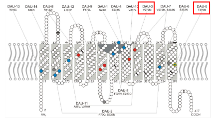
The RhD protein is a 12-pass transmembrane glycoprotein encoded by the RHD gene (Figure 5). The allelic diversity of the RHD locus gives rise to variant forms that may express an apparently normal D antigen with reduced surface density, termed weak D, or altered epitopes that can elicit an allo-immune response, termed partial D. Weak D is often associated with alterations in mRNA processing/stability, or with amino acid substitutions in transmembrane or intracellular domains. In contrast, partial D variants more frequently involve amino acid substitutions in one of the six extracellular loops, or truncation of the protein resulting in epitope loss (1). While this topographical distinction is intuitive, the underlying biochemistry is likely more complex due to the rare instances of anti-D formation in individuals with weak D genotypes. Additionally, the subtle shifts in protein architecture translate into clinical uncertainty regarding alloimmunization risk.
The risk of RhD alloimmunization and associated complications is well defined in RhD-negative individuals. Yazer et al. (2007) reported an alloimmunization rate of 22% among RhD-negative patients who received emergency RhD-positive transfusions. In this population, anti-D alloantibodies are generally IgG, reactive at 37 °C, non-complement fixing, and capable of causing extravascular hemolysis of transfused RhD-positive RBCs. Rarely, IgM anti-D antibodies develop and fix complement, leading to intravascular hemolysis. In females of childbearing potential, Rh alloimmunization is a significant risk factor for hemolytic disease of the fetus and newborn (HDFN), where maternal IgG anti-D can cross the placenta and target fetal erythrocytes, potentially resulting in complications including fetal anemia, hydrops fetalis, and kernicterus. The alloimmunization risk associated with RhD variants is less well established, and the limitations of conventional clinical laboratory testing make their detection and classification challenging.
An individual’s RhD status is a core feature of their serologically derived blood type, and a standard component of the commonly ordered type and screen. RhD typing is typically performed at the immediate spin (IS) phase using a monoclonal IgM anti-D reagent that recognizes multiple RhD epitopes, and the degree of agglutination serves as the measure of positivity (on a scale from 0 to 4+). The so-called ‘weak D’ test extends testing to the AHG (anti-human globulin or Coombs) phase. The common assumption is that individuals with weak D variants will initially type as RhD-negative but may become positive on the ‘weak D’ test, while those with partial D variants will type as RhD-positive. Although this is true with some variants, many fall outside of this expected pattern. Therefore, in practice, serologic testing alone cannot differentiate between weak and partial D phenotypes due to their variable presentations. When serologic testing suggests a weak or variant D, particularly in the setting of weak agglutination with the RhD typing reagent, or discrepant results between different typing methods, a descriptive interpretation of ‘serologic weak D’ may be used and further investigation by molecular testing may be warranted (7, 8).
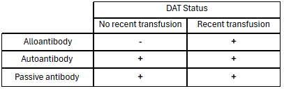
There are several different possibilities to consider when evaluating a patient whose RBCs type as RhD positive but also has an apparent anti-D antibody in their plasma. The DAT (direct antiglobulin or Coombs test) helps determine whether antibodies are bound to RBCs in circulation, though its interpretation is highly dependent on the clinical context. Notably, the DAT cannot distinguish between antibodies bound to host RBCs or foreign RBCs, and its results must be interpreted with caution in the setting of recent RBC transfusion. In an RhD-positive patient with an anti-D antibody, a negative DAT would suggest that the patient has a partial D antigen and the anti-D is an alloantibody against the portion of the D antigen which the patient lacks. In this setting, the allo anti-D antibody would not bind to the patient’s own RBCs. If the patient was recently transfused with RhD positive RBCs, the DAT may be positive because the antibody is binding to the transfused RhD-positive RBCs. Other possible explanations for a positive DAT in this setting include a warm auto antibody with an anti-D-like specificity, or a passive anti-D antibody, for example, due to Rh immune globulin (RhIG) administration for treatment of immune thrombocytopenic purpura (ITP), or in the setting of a transplant complicated by Passenger Lymphocyte Syndrome where the donor had an anti-D antibody and the recipient was RhD positive. Autoantibodies binding to a patient’s own RBCs can only be definitively inferred when no foreign RBCs or antibodies are present (Table 1). In cases where alloantibodies are identified against antigens that appear to be present on the patient’s own RBCs by serological testing, and the DAT is negative, additional molecular genotyping is suggested.
Here, we report a case of a pediatric patient with SCD who was typed as RhD-positive (Figure 1) but developed an anti-D antibody following transfusion with RhD-positive RBC units (Figure 2). DAT negativity with the newly identified antibody suggested alloantibody formation (Figure 3). The patient’s most recent transfusion with two units of RBCs, one RhD positive and the other RhD negative, occurred about a month prior to their presentation. Laboratory markers, including hemoglobin, hematocrit and % hemoglobin S (Figure 4) were suggestive of hemolysis of the recently transfused RhD positive RBC unit. The identification of anti-D in this RhD-positive patient prompted molecular testing, which revealed the presence of a DAU0/DAU3 allele combination.
DAU0 and DAU3 belong to the DAU cluster of RhD variants (Figure 5), which represents a group of related alleles that all harbor the Thr379Met amino acid substitution (9). The DAU0 allele, which has a relatively high allele frequency in populations of African descent (16%), is considered the “primordial” allele because it carries only the cluster-defining mutation (9). Other DAU variants, including DAU3, harbor additional mutations that can alter protein expression or epitope structure (9). Although DAU0 is generally considered a weak D variant with low immunogenic potential due to presentation of normal epitopes, instances of alloimmunization have been identified (9-11). Takasaki et al. (2023) reported anti-D in 5.4% (5/92) of patients with DAU0 and reduced RBC survival in 30% of cases. DAU3, in contrast, is suspected to alter surface epitopes and has been linked to anti-D formation, although one reported case was determined to be an autoantibody (10,11). Given the rarity of RhD variants and the limited data on clinical outcomes, defining the risk of alloimmunization and the associated clinical impact remains difficult.
When considering the optimal transfusion strategy for patients with an RhD variant, with or without an anti-D antibody, the decision ultimately comes down to risk management and logistical feasibility. Weak D types 1, 2 and 3 are well-described in the literature and patients with these variants do not typically make clinically significant allo anti-D antibodies; therefore, patients with weak D types 1, 2 and 3 are generally interpreted, and clinically managed, as RhD positive for the purposes of transfusion and RhIG prophylaxis during pregnancy (7, 8). Some authors have recommended the use of RhD-positive units for certain other weak D examples, particularly in settings where RhD-negative blood is limited (7). While this approach may be reasonable in some cases, a more conservative strategy may be preferred when limited information regarding clinical significance is available in the literature, especially when treating chronically transfused patients with heightened risk of alloimmunization. In such patients, RhD-negative transfusions can mitigate the risk of immune-mediated hemolysis and potential impact on transfused RBC lifespan. There are challenging situations in which patients present with multiple alloantibodies, or antibodies directed against high-frequency antigens, which may significantly limit availability of antigen-negative RBCs to meet their needs. In these cases, a trial of RhD-positive blood may be necessary due to inventory constraints. Ultimately, however, transfusion decisions must be individualized, balancing risk against product availability. In our case, given the patient’s currently known requirements, antigen-negative product availability is sufficient for utilization of RhD negative RBCs in all future transfusions, and that is the approach that will be taken until further information is available regarding the clinical significance of this allo anti-D antibody in a patient with the RHD*DAU0/DAU3 variant.
Citations
- Avent, N. D., & Reid, M. E. (n.d.). The Rh blood group system: a review.
- Chou, S. T., Alsawas, M., Fasano, R. M., Field, J. J., Hendrickson, J. E., Howard, J., Kameka, M., Kwiatkowski, J. L., Pirenne, F., Shi, P. A., Stowell, S. R., Thein, S. L., Westhoff, C. M., Wong, T. E., & Akl, E. A. (2020). American society of hematology 2020 guidelines for sickle cell disease: Transfusion support. In Blood Advances (Vol. 4, Issue 2, pp. 327–355). American Society of Hematology. https://doi.org/10.1182/BLOODADVANCES.2019001143
- Chou, S. T., Evans, P., Vege, S., Coleman, S. L., Friedman, D. F., Keller, M., & Westhoff, C. M. (2018). RH genotype matching for transfusion support in sickle cell disease. In Blood (Vol. 132, Issue 11). http://ashpublications.org/blood/article-pdf/132/11/1198/1372747/blood851360.pdf
- Chou, S. T., Jackson, T., Vege, S., Smith-Whitley, K., Friedman, D. F., & Westhoff, C. M. (2013). High prevalence of red blood cell alloimmunization in sickle cell disease despite transfusion from Rh-matched minority donors. Blood, 122(6), 1062–1071. https://doi.org/10.1182/blood-2013-03-490623
- Daniels, G. (2013). Variants of RhD - current testing and clinical consequences. In British Journal of Haematology (Vol. 161, Issue 4, pp. 461–470). https://doi.org/10.1111/bjh.12275
- Fasano, R. M., Meyer, E. K., Branscomb, J., White, M. S., Gibson, R. W., & Eckman, J. R. (2019). Impact of Red Blood Cell Antigen Matching on Alloimmunization and Transfusion Complications in Patients with Sickle Cell Disease: A Systematic Review. In Transfusion Medicine Reviews (Vol. 33, Issue 1, pp. 12–23). W.B. Saunders. https://doi.org/10.1016/j.tmrv.2018.07.003
- Flegel, W. A., Denomme, G. A., Queenan, J. T., Johnson, S. T., Keller, M. A., Westhoff, C. M., Katz, L. M., Delaney, M., Vassallo, R. R., Simon, C. D., & Sandler, S. G. (2020). It’s time to phase out “serologic weak D phenotype” and resolve D types with RHD genotyping including weak D type 4. In Transfusion (Vol. 60, Issue 4, pp. 855–859). Blackwell Publishing Inc. https://doi.org/10.1111/trf.15741
- Sandler, S. G., Flegel, W. A., Westhoff, C. M., Denomme, G. A., Delaney, M., Keller, M. A., Johnson, S. T., Katz, L., Queenan, J. T., Vassallo, R. R., & Simon, C. D. (2015). It’s time to phase in RHD genotyping for patients with a serologic weak D phenotype. In Transfusion (Vol. 55, Issue 3, pp. 680–689). https://doi.org/10.1111/trf.12941
- Srivastava, K., Polin, H., Sheldon, S. L., Wagner, F. F., Grabmer, C., Gabriel, C., Denomme, G. A., & Flegel, W. A. (2016). The DAU cluster: a comparative analysis of 18 RHD alleles, some forming partial D antigens. Transfusion, 56(10), 2520–2531. https://doi.org/10.1111/trf.13739
- Takasaki, K., Friedman, D. F., Uter, S., Vege, S., Westhoff, C. M., & Chou, S. T. (2023). Variant RHD alleles and Rh immunization in patients with sickle cell disease. British Journal of Haematology, 201(6), 1220–1228. https://doi.org/10.1111/bjh.18774
- Wagner, F. F., Ladewig, B., Angert, K. S., Heymann, G. A., Eicher, N. L., & Flegel, W. A. (2002). The DAU allele cluster of the RHD gene. Blood, 100(1), 306–311. https://doi.org/10.1182/blood-2002-01-0320
- Yazer, M. H., & Triulzi, D. J. (2007). Detection of anti-D in D- recipients transfused with D+ red blood cells. Transfusion, 47(12), 2197–2201. https://doi.org/10.1111/j.1537-2995.2007.01446.x
- Teles, B., Santos, T., & Castilho, L. (2023). Risk of clinically significant anti-D in Sickle Cell Disease patients with partial D transfused with D-positive red blood cells Vox Sanguinis, 118(S1), 263. https://doi.org/10.1111/vox.13433_1