
Final Diagnosis
The patient’s bone marrow dysplasia, ring sideroblasts, and SF3B1 mutation together were diagnostic of a myelodysplastic neoplasm (MDS) which was best classified as MDS with mutated SF3B1 (International Consensus Classification) / MDS with low blasts and SF3B1 mutation (WHO 5th Edition). In addition, the abnormal T-cell population combined with the presence of the STAT3 mutation was consistent with a diagnosis of T-cell large granular lymphocytic leukemia (T-LGLL) (ICC & WHO 5th Edition).
Discussion
1. MDS with Mutated SF3B1 (ICC) / MDS with Low Blasts and SF3B1 Mutation (WHO 5th Edition):
Myelodysplastic neoplasms (MDS) are clonal hematopoietic stem cell neoplasms characterized by cytopenia(s) and morphologic dysplasia of myeloid lineage cells. MDS tends to present in older adults, and its rate progressively increases with age [1,2].
In general, a diagnosis of MDS requires the following [1,2]:
1. Cytopenia in at least 1 myeloid lineage:
a. Hemoglobin (Hgb) <13 g/dL for males & <12 g/dL for females
b. Absolute neutrophil count (ANC) <1.8 x 10⁹/L
c. Platelets (Plt) <150 x 109/L
2. Dysplasia in at least 1 myeloid lineage:
a. Erythroid, granulocytic, and/or megakaryocytic lineages
i. A dysplastic lineage requires dysplasia in ≥10% of cells of that lineage
ii. The dysplastic lineage(s) can differ from the cytopenic lineage(s)
3. Evaluation of the percentage of blasts in the bone marrow and peripheral blood:
a. Thresholds vary for each MDS subtype
i. Although, ≥20% blasts immediately excludes MDS of any subtype
b. For MDS-SF3B1, specifically:
i. <5% blasts in the bone marrow
ii. <2% blasts in the peripheral blood
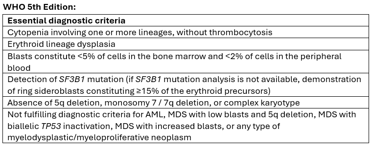
The presence of ring sideroblasts in the context of MDS can serve as a clue to a more refined diagnosis of MDS with mutated SF3B1. In such cases, confirmation of an SF3B1 mutation via molecular testing is required for diagnosis (VAF ≥10%). However, if molecular testing is not available, the WHO 5th Edition classification permits ring sideroblasts to serve as a surrogate in making this diagnosis if the amount of ring sideroblasts constitutes ≥15% of erythroid precursors. It is important to note that ring sideroblasts can be seen in various non-neoplastic conditions, including alcohol, toxins (e.g. lead, benzene), drugs (e.g. isoniazid), copper deficiency, and congenital sideroblastic anemia, and therefore, these conditions should be excluded [2,3].
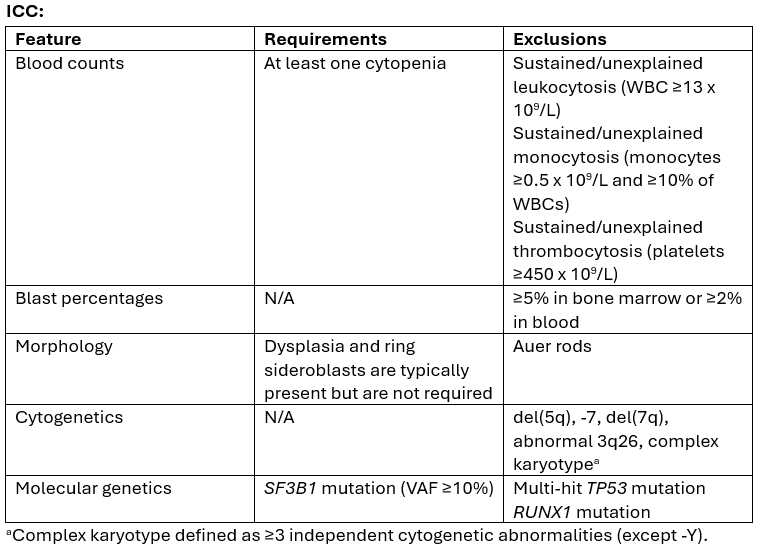
Finally, before making a diagnosis of MDS with mutated SF3B1, exclusion of specific features is necessary. Some of these features suggest an alternative MDS subtype and/or negate the favorable prognosis associated with the SF3B1 mutation. These features include: del(5q), -7/del(7q), inv(3), abnormal 3q26, complex karyotype (≥3 independent cytogenetic abnormalities, except -Y), multi-hit TP53 mutation, and RUNX1 mutation [3].
Notably, MDS with mutated SF3B1 carries the best prognosis among all MDS subtypes [1,4,5]. Ring sideroblasts do not have a prognostic significance independent of the SF3B1 mutation [3].
The full diagnostic criteria specific to both the ICC and WHO 5th Edition classification systems are summarized (adapted) below [2,3]:
2. T-Cell Large Granular Lymphocytic Leukemia (T-LGLL) (ICC & WHO 5th Edition):
T-LGLL is a clonal proliferation of cytotoxic T cells that presents in older adults, and due to its indolent nature, does not always produce symptoms. Notably, one third of patients are asymptomatic. For patients who do experience symptoms, their clinical presentation is often related to underlying cytopenia(s). T-LGLL is also frequently associated with autoimmune disorders, including rheumatoid arthritis, systemic lupus erythematosus, and Sjögren syndrome [6,7].
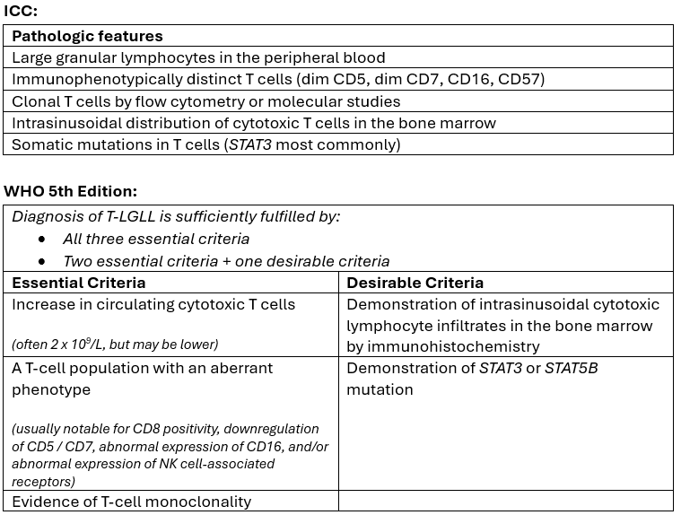
Flow cytometric studies on the peripheral blood demonstrate an expanded CD3+ CD8+ TCRαβ+ T-LGLL population additional features of a mature effector memory phenotype (CD57+, CD45RA+, CD62-). Alternatively, a minority of cases express CD4 (alone or in combination with dim/partial CD8), and less than 10% of cases are of TCRγδ T-cell lineage. The neoplastic cells often express TIA1, perforin, granzyme B, and/or granzyme M, which helps support their cytotoxic profile. Expression of NK cell-associated antigens (CD16+57) is frequent, but not disease-specific [6,7].
The pathologic features specific to both the ICC and WHO 5th Edition classification systems are summarized (adapted) below [6,7]:
Sources
- Hasserjian RP. Introduction to Premalignant Clonal Cytopenias and Myelodysplastic Syndromes. In: Arber DA, Borowitz MJ, Cook JR, de Leval L, Goodlad JR, Hasserjian RP, King RL, Kvasnicka HM, Orazi A, editors. The International Consensus Classification of Myeloid and Lymphoid Neoplasms. 1st ed. Philadelphia (PA): Lippincott Williams & Wilkins; 2025. p. 234-238.
- Fontenay M, Germing U, Nogueira Zerbini MC, Ito M, Khoury JD, Naresh KN, Nardi V, Park S, Ades L, Haase D, Jansen JH, Ogawa S, Sallman D, Patkar N, Platzbecker U, Komrokji R. Myelodysplastic neoplasm with low blasts and SF3B1 mutation. In: WHO Classification of Tumours Editorial Board. Haematolymphoid tumours [Internet]. Lyon (France): International Agency for Research on Cancer; 2024 [cited 2025 Nov. 17]. (WHO classification of tumours series, 5th ed.; vol. 11). Available from: https://tumourclassification.iarc.who.int/chaptercontent/63/23.
- Siddon AJ, Hellström-Lindberg E, Malcovati L. Myelodysplastic Syndrome with Mutated SF3B1. In: Arber DA, Borowitz MJ, Cook JR, de Leval L, Goodlad JR, Hasserjian RP, King RL, Kvasnicka HM, Orazi A, editors. The International Consensus Classification of Myeloid and Lymphoid Neoplasms. 1st ed. Philadelphia (PA): Lippincott Williams & Wilkins; 2025. p. 247-252.
- Ball S, Singh AM, Al Ali N, Aguirre LE, Jain AG, Schwabkey ZI, Tinsley-Vance SM, Chan O, Kuykendall A, Sweet K, Lancet JE, Padron E, Sallman DA, Komrokji RS; A product of “clash of titans" or true reflection of disease biology? Validation of 2022 WHO and ICC classifications in a large dataset of patients with myelodysplastic syndrome. Blood Nov. 2022; 140 (Supplement 1): 1118–1120. doi: https://doi.org/10.1182/blood-2022-170158.
- Malcovati L, Karimi M, Papaemmanuil E, Ambaglio I, Jädersten M, Jansson M, Elena C, Gallì A, Walldin G, Della Porta MG, Raaschou-Jensen K, Travaglino E, Kallenbach K, Pietra D, Ljungström V, Conte S, Boveri E, Invernizzi R, Rosenquist R, Campbell PJ, Cazzola M, Hellström Lindberg E. SF3B1 mutation identifies a distinct subset of myelodysplastic syndrome with ring sideroblasts. Blood. 2015 Jul 9;126(2):233-41. doi: 10.1182/blood-2015-03-633537. Epub 2015 May 8. PMID: 25957392; PMCID: PMC4528082.
- Morice WG, Cheng CL, Loughran TPJ, Gujral S, Naresh KN, Lamy T, Semenzato G. T-large granular lymphocytic leukaemia. In: WHO Classification of Tumours Editorial Board. Haematolymphoid tumours [Internet]. Lyon (France): International Agency for Research on Cancer; 2024 [cited 2025 Nov. 17]. (WHO classification of tumours series, 5th ed.; vol. 11). Available from: https://tumourclassification.iarc.who.int/chaptercontent/63/210.
- Hsi ED, Maciejewski JP, Wang SA. T-Cell Large Granular Lymphocytic Leukemia. In: Arber DA, Borowitz MJ, Cook JR, de Leval L, Goodlad JR, Hasserjian RP, King RL, Kvasnicka HM, Orazi A, editors. The International Consensus Classification of Myeloid and Lymphoid Neoplasms. 1st ed. Philadelphia (PA): Lippincott Williams & Wilkins; 2025. p. 970-978.