
Final Diagnosis
Uterine Carcinosarcoma
Discussion
Carcinosarcoma of the uterus, previously termed malignant mixed Müllerian tumor, is a rare endometrial malignancy characterized by a biphasic growth pattern consisting of both carcinomatous and sarcomatous elements. The prevalence of these malignancies has increased over the last several decades, comprising approximately 5.7% of endometrial cancers in 2016 compared with 1.7% in 2000 (6). Additionally, there has been a shift in patient demographics. Although the historical peak incidence has been among patients 70–79 years of age, the largest interval increase in recent years has occurred in the 60–69-year age group (5). As such, carcinosarcoma is largely considered a disease of older individuals, and its occurrence in a patient in their third decade of life, as in the present case, is exceedingly uncommon, although not absent from the literature (2-4).
The pathogenesis of carcinosarcoma is not entirely understood. However, current evidence supports a metaplastic or dedifferentiation process in which the sarcomatous component arises from the high-grade carcinomatous component through epithelial-to-mesenchymal transition (1). This contrasts with the historical concept of two independent neoplastic processes arising simultaneously within the epithelium and stroma. In terms of Bokhman’s classification, carcinosarcoma falls within the type II endometrial cancer category, reflecting its largely estrogen-independent development (6).
Although carcinosarcoma is considered a high-grade neoplasm, the carcinomatous component can vary morphologically in grade and differentiation. The sarcomatous component may be homologous, with elements resembling tissues native to the uterus, such as endometrial stromal sarcoma, leiomyosarcoma, or fibrosarcoma, or heterologous, with elements resembling tissues not normally present in the uterus, including rhabdomyosarcoma, chondrosarcoma, and osteosarcoma (6). In the present case, although some areas showed features more consistent with homologous stromal differentiation, a substantial component demonstrated prominent hyalinization with cartilaginous-appearing areas (Figure 3). While these findings are consistent with heterologous cartilaginous differentiation, chondroid matrix has also been described in other neoplasms such as corded and hyalinized endometrioid carcinoma (CHEC) (8, 9).
Immunophenotypically, the staining pattern of carcinosarcoma reflects its biphasic nature. The epithelial component is generally positive for keratins, including AE1/AE3 and CAM5.2, as well as EMA and PAX8. The stromal component is generally positive for vimentin and may show focal expression of keratins and EMA. Heterologous elements demonstrate expression of lineage-specific markers, such as desmin, myogenin, and MyoD1 in rhabdomyoblastic differentiation and S100 in cartilaginous differentiation (6). In the present case, S100 showed patchy positivity within the non-glandular component, supporting cartilaginous differentiation.
Given the prominent stromal hyalinization and cording of tumor cells, an important diagnostic consideration is CHEC, an entity first described by Murray et al. in 2005 (12). Like carcinosarcoma, CHEC may have a biphasic appearance, however, conventional CHEC is typically composed of relatively bland epithelioid or spindled cells arranged in cords within a hyalinized stroma and associated with low-grade endometrioid carcinoma (12). Stromal features may include myxoid, osteoid, and, less commonly, chondroid matrix (8, 9). Conventionally, CHEC has demonstrated clinical behavior similar to that of low-grade endometrioid carcinoma and is often treated as such.
Several features have traditionally been useful in distinguishing CHEC from carcinosarcoma. Conventional CHEC is generally low-grade, therefore, high-grade features favor carcinosarcoma. A characteristic immunophenotypic feature of CHEC is nuclear β-catenin expression, which has been identified in approximately 92% of reported cases and corresponds to CTNNB1 alterations in a substantial proportion of cases (10, 11). Hormone receptor expression is generally retained within the glandular component but may be reduced or lost in the corded component. In the present case, the high-grade features, focal areas with more conventional carcinosarcoma morphology (Figure 5), and predominantly membranous/cytoplasmic β-catenin expression favor a diagnosis of carcinosarcoma.
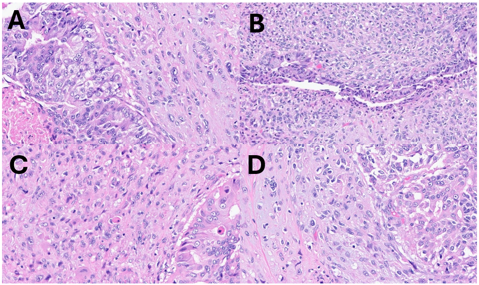
More recently, however, a small number of CHECs with high-grade features have been reported, further complicating the distinction between these entities. Travaglino et al. described six high-grade CHECs, five of which demonstrated either MMR-deficient or p53-abnormal molecular signatures. These tumors also exhibited morphologic features overlapping with carcinosarcoma, including marked cytologic atypia, necrosis, and osteoid or myxoid/chondromyxoid matrix (8, 9). Additionally, only two of the six cases demonstrated nuclear β-catenin expression, and none harbored a hotspot CTNNB1 alteration. Taken together, these findings suggest that some of the features traditionally used to distinguish CHEC from carcinosarcoma may be less specific in the setting of high-grade morphology than previously thought (8, 9).
Overall, the morphologic and immunophenotypic findings in the present case favor carcinosarcoma, particularly given the high-grade cytologic features and focal areas of more conventional carcinosarcoma morphology. However, the prominent hyalinized stroma, corded growth pattern, and cartilaginous-appearing matrix show morphologic overlap with the recently described high-grade CHEC. While the findings in the present case are most consistent with carcinosarcoma, CHEC with high-grade features remains a diagnostic consideration.
Citations
- Pillai, M. S. G. et al. Uterine carcinosarcoma: Unraveling the role of epithelial‐to‐mesenchymal transition in progression and therapeutic potential. FASEB J. 38, e70132–e70132 (2024).
- FUJIWARA, H. et al. Unsuspected Uterine Carcinosarcoma (heterologous) Diagnosed following Conservative Therapies with Medroxyprogesterone Acetate for Presumed Early‐Stage Endometrial Carcinoma. Am. J. Reprod. Immunol. 47, 129–131 (2002).
- Wei, N., Bai, S., Kong, D., Wang, X. & Mi, X. Endometrial carcinosarcoma in a young female with absence of established risk factors: a case report. Front. Oncol. 15, 1656841 (2025).
- Iida, Y. et al. A rare case of uterine carcinosarcoma in a 21-year-old woman with hereditary breast and ovarian cancer syndrome: A case report. Oncol. Lett. 31, 194 (2026).
- Matsuo, K., Ross, M. S., Machida, H., Blake, E. A. & Roman, L. D. Trends of uterine carcinosarcoma in the United States. J. Gynecol. Oncol. 29, e22 (2017).
- Matsuzaki, S. et al. Uterine carcinosarcoma: Contemporary clinical summary, molecular updates, and future research opportunity. Gynecol. Oncol. 160, 586–601 (2021).
- Zhang, X., Cui, F., Yuan, Y., Cheng, H. & Cao, Z. High-grade corded and hyalinized endometrioid carcinoma with high microsatellite instability and STK11 mutation: A case report and literature review. Oncol. Lett. 30, 389 (2025).
- Travaglino, A. et al. Corded and hyalinized endometrioid endometrial carcinoma with high-grade features: a clinicopathological and TCGA-based molecular analysis. Virchows Arch. 482, 671–678 (2023).
- Travaglino, A. et al. Corded and hyalinized endometrioid carcinoma: Summary of clinical, histological, immunohistochemical and molecular data. Pathol. - Res. Pract. 247, 154515 (2023).
- Safdar, N. S. et al. Corded and Hyalinized and Spindled Endometrioid Endometrial Carcinoma. Am. J. Surg. Pathol. 45, 1038–1046 (2021).
- Ladwig, N. R., Umetsu, S. E., Zaloudek, C., Rabban, J. & Garg, K. Corded and Hyalinized Endometrioid Adenocarcinoma (CHEC) of the Uterine Corpus are Characterized by CTNNB1 Mutations and Can Show Adverse Clinical Outcomes. Int. J. Gynecol. Pathol. 40, 103–115 (2021).
- Murray, S. K., Clement, P. B. & Young, R. H. Endometrioid Carcinomas of the Uterine Corpus With Sex Cord-like Formations, Hyalinization, and Other Unusual Morphologic Features. Am. J. Surg. Pathol. 29, 157–166 (2005).