Authors: Nathaniel Bigot, DO, Suzanne Bakdash, MD, MPH
Case Presentation
A pediatric patient with sickle cell disease (SCD) presented to our SCD clinic for routine follow-up and pre-transfusion testing. Cerebrovascular screening by transcranial Doppler (TCD) consistently showed elevated peak systolic flow in the middle and anterior cerebral arteries, indicating a high risk for pediatric stroke. To mitigate this risk, the patient undergoes monthly partial exchange transfusions to maintain hemoglobin S (HbS) levels below 35%. The patient types as group O RhD-positive with a negative antibody screen, though anti-Fya antibody was detected in the past. In accordance with current American Society of Hematology (ASH) guidelines concerning transfusion support for alloimmunized patients with SCD (2), red blood cell (RBC) units were selected to match the patient’s Rh (D, C/c, E/e), Kell (K), Duffy (Fya), Kidd (Jkb), and S/s phenotype. Due to these matching requirements, both group O Rh-positive and Rh-negative units were used for exchange transfusion, the most recent of which occurred one month prior to presentation.
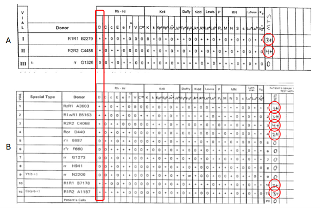
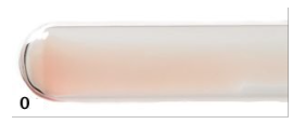
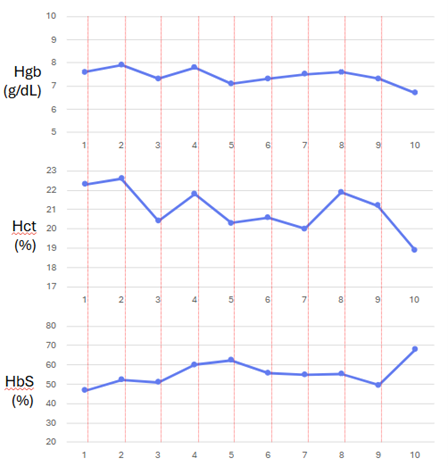
On current pre-transfusion testing, the patient was again typed as group O RhD-positive (Figure 1), but the antibody screen revealed a new anti-D antibody (Figure 2). The DAT (direct antiglobulin or Coombs test) was negative (Figure 3). Laboratory evaluation showed a mild drop in hemoglobin and hematocrit, together with an increase in HbS percentage. These findings fell outside the patient’s historical pre-transfusion baseline and suggested possible hemolysis with bone marrow compensation (Figure 4). The suspected hemolysis, together with the discrepancy between the blood type and newly identified anti-D antibody, prompted the blood bank to provide exclusively RhD-negative, phenotypically matched RBC units to minimize the risk of future hemolytic transfusion reactions. A blood sample was sent to a reference laboratory for RhD genotyping, which identified RHD*DAU0/DAU3.