
Authors: Vandana Panakkal, M.D and Alesia Kaplan, M.D.
Date of Publication: January 1, 2026
Case Scenario
A female in her mid-20s, presented for antenatal care at 8 weeks of gestational age. Her past obstetric history included a spontaneous first trimester-miscarriage managed with dilation and curettage. Laboratory evaluation excluded fetal chromosomal abnormalities and maternal thrombophilia. Blood grouping showed an ABO discrepancy with unexpected antibody reactivity in reverse typing. Antibody screening was positive while the direct antiglobulin test (DAT) was negative. On antibody identification by tube testing, patient's plasma reacted with all reagent RBCs at immediate spin (2+), demonstrated weak reactivity at 37 °C, and was strongly positive (3+) in the antihuman globulin (AHG) phase, with a negative auto control. These findings suggested the presence of an alloantibody directed against a high-frequency antigen. Enzyme-treated reagent red cells exhibited enhanced reactivity with patient plasma at 37 °C (4+) and in the AHG phase (4+) confirm that the antibody is directed against an enzyme-resistant antigen. After treatment with 0.01 M dithiothreitol (DTT), plasma showed no reactivity at 37 °C and reduced reactivity (2+) in the AHG phase, indicating that the antibody contained both IgG and IgM components. Testing of patient plasma against rare PP1Pk-negative cells revealed no reactivity, and the patient’s RBCs typed negative, when tested with reference plasma containing anti-PP1Pk antibodies. ABO/Rh typing using adsorbed plasma confirmed patient’s blood group as A positive. One year later, she conceived again. Given her history of anti-PP1Pk alloimmunization, antibody titers were closely monitored, and paternal antigen testing showed an O-positive, PP1Pk/P1 positive blood type. This pregnancy was successfully managed with therapeutic plasma exchange, intravenous immunoglobulin (IVIG), and serial monitoring of anti-PP1Pk titers, culminating in a vaginal delivery of a healthy female infant at 38 weeks and 3 days. Management of this pregnancy was reported previously (4).
At the initial visit during the current pregnancy, blood grouping, antibody screening, and antibody identification were repeated using the methods described above. Presence of anti-PP1Pk antibody was confirmed, with an initial titer of 1:32. Therapeutic plasmapheresis (1.5 times the estimated total plasma volume, approximately 3,750 mL, with human albumin replacement) was initiated twice weekly beginning at the 10th week of gestation. In addition, intravenous immunoglobulin (IVIG) was administered weekly at a dose of 1 g/kg body weight (100 g), commencing at the 9th week of gestation (Figure 1A). Anti-PP1Pk titers were monitored at least once a week and maintained below 1:32 throughout the pregnancy (Figure 1B). Beginning at 16 weeks of gestation, weekly fetal Doppler imaging was performed with estimation of middle cerebral artery peak systolic velocity (MCA-PSV) to detect fetal anemia. Fetal non-stress testing (NST) was scheduled to begin at 24 weeks in conjunction with plasmapheresis. However, it was not performed due to patient preference.
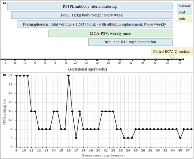
Maternal anemia was managed with intravenous iron sucrose (200mg x 3 doses) and intramuscular vitamin B12 (1000 mcg) around 25 weeks of gestation along with oral supplementation. Blood draws were minimized to reduce transfusion requirements. A directed donation of a PP1Pk-negative RBC unit from a relative was attempted but unsuccessful; the blood bank maintained PP1Pk-negative units in frozen inventory, and one unit was thawed for delivery as a precaution.
At 38 weeks and 4 days of gestation, the patient underwent a planned external cephalic version for breech presentation. Following an unsuccessful attempt, an emergent cesarean section was performed, resulting in the delivery of a healthy female neonate. Intraoperative cell salvage was employed to collect autologous RBCs. Overall blood loss was within expected limits, and no peripartum blood product transfusion was required. The neonate was blood group A positive, with a negative direct antiglobulin test. The postpartum course was uneventful, and both mother and neonate were well at outpatient follow-up.