
Authors: Pranav P. Patwardhan1, Maria Grupillo1, Cody Marshall2, Simon I. Chiosea1
Date of Publication: April 1, 2026
1Department of Pathology, University of Pittsburgh Medical Center, Pittsburgh, PA
2Department of Pathology, UPMC Northern Regional Hospital
Case History
A female patient in her eighties presented to the clinic with complaints of persistent diarrhea. She also documented around 15-pound weight loss in the recent past and was a non-smoker. Subsequent imaging studies (CT scan) demonstrated bilateral lung nodules and a 4.6 x 4.5 cm right hepatic lobe mass. A liver biopsy was performed, and material was sent for histopathological examination and molecular genomic testing (solid tumor Next generation sequencing studies)
Histopathologic Findings and Molecular Studies
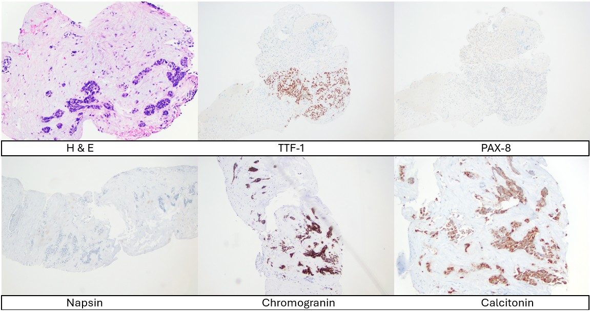
Histopathologic examination of the liver biopsy demonstrated findings diagnostic for involvement by a metastasis. Preliminary immunohistochemical evaluation demonstrated the tumor to be positive for CK7 and TTF-1 and negative for CK20, CDX2, GATA-3 and PAX-8 (See figure 1). Thus, a provisional diagnosis of metastatic adenocarcinoma, favoring lung primary was favored.
Concurrently, solid tumor next generation sequencing using UPMC Oncomine comprehensive assay was performed on the tumor tissue. A pathogenic RET deletion (p.D898_E901del) was detected at an allele fraction of 23% (See figure 2). Variants of uncertain significance were detected in BRCA2 and CREBBP genes. These molecular pathology results prompted further evaluation and immunohistochemical workup of the liver biopsy.
Subsequent immunohistochemical stains demonstrated the tumor cells to be positive for calcitonin, CEA, synaptophysin, chromogranin and negative for napsin A (See figure 1). The proliferation index on Ki67 was <5%.
RNA-seq performed was negative.
Concurrently, ultrasound studies performed on the thyroid revealed a 17 x 13 x 12 mm, TIRADS-5 hypoechoic, interpolar mass with tiny hyperechoic, calcific foci in the right thyroid lobe. PET-CT performed also revealed PET avidity in the posterior right thyroid nodule, high right paratracheal region, bilateral pulmonary metastasis and in the hepatic mass.
Thus, based on the additional immunohistochemical results, a pathogenic RET deletion detected on NGS testing and supporting additional imaging findings, a diagnosis of metastatic medullary thyroid carcinoma was made and communicated to the clinical team.
Plasma circulating tumor DNA testing (Guardant360) also identified a RET D898_E901 deletion at VAF of 8.9%.

Clinical Follow Up
Initially, a lung non-small cell carcinoma directed chemotherapy was planned, given the initial impression favoring a lung primary. However, with the diagnosis of metastatic medullary thyroid carcinoma and detection of a pathogenic RET deletion, the patient was advised selpercatinib. A Blood calcitonin level was performed which was high (219 pg/ml). A germline mutation panel screening was negative. Subsequent to the receipt of selpercatinib, the serum calcitonin levels dropped to 51.7 pg/ml and <2 pg/ml before selpercatinib had to be discontinued due to side effects which included weakness in the legs, elevated liver enzymes and elevated blood pressure. On last available follow-up, the clinical plan was to restart a lower dose of selpercatinib, when the clinical conditions would allow. Owing to the discontinuation of therapy, the blood calcitonin level on the last follow up was increased (219 pg/ml).
Discussion
We present this case as it demonstrates the utility of molecular genomic testing in the correct diagnosis and subsequent therapy in solid tumor malignancies. There are also some useful teaching points in this case to highlight and discuss.
First, it demonstrates the importance of evaluating histopathology and other ancillary studies in the context of clinical history, imaging findings and other laboratory tests. It is always a helpful idea to keep a broad differential diagnosis in the initial stages of case evaluation to not miss some outlier cases.
Secondly, it highlights the importance of knowledge of immunohistochemical expression in different tissues and pathologies. Thyroid transcription factor-1 (TTF-1) is expressed in thyroid follicular cells, in the lung and can also be variably expressed in gynecologic tissues 1,2. On a similar note, PAX-8 immunohistochemical stain highlights thyroid follicular cells, renal parenchyma and is positive in gynecologic tissues. PAX-8 is positive in most differentiated thyroid neoplasms (papillary and follicular type) but it’s staining is variable and inconsistent in medullary thyroid carcinoma 3. Thus, using a panel of immunostains is helpful in the context of evaluation of neoplasms to avoid these pitfalls. The liver metastasis in this case was positive for TTF-1 and negative for PAX-8 raising the possibility of lung adenocarcinoma, especially in the context of clinical findings with lung lesions and large liver metastasis. Subsequent Napsin-A was negative and calcitonin, synaptophysin, chromogranin were positive, confirming a diagnosis of medullary thyroid carcinoma in this case.
Finally, this case also demonstrates the utility of molecular genomic sequencing in such cases, where it may help by identifying pathognomic or diagnostic genomic alterations. Medullary thyroid carcinomas are associated with germline and somatic RET mutations. RET mutations are also associated with a more aggressive disease4. RET D898_E901 deletion is an in-frame oncogenic deletion seen in exon 15 of RET kinase domain and has been described in medullary thyroid carcinoma5. It is a gain-of-function mutation and thus can be targeted with RET-targeted therapy. In this case, the solid tumor NGS panel identified this deletion. On further literature review, it was found to be associated with medullary thyroid carcinoma and not definitively with other solid tumors, including lung adenocarcinomas, which can show RET fusions in a subset of cases. This prompted additional immunohistochemical workup in this case which led to a diagnosis of medullary thyroid carcinoma. The blood calcitonin levels dropped with initiation of targeted therapy, practically demonstrating the clinical utility of detection of a targetable genetic alteration.
Thus, molecular genomic testing potentially helped in the accurate diagnosis and selection of therapy in this case, and we hope this case demonstrates the utility of next generation sequencing in the evaluation of metastatic lesions.
References
- Möller K et al. TTF-1 is a highly sensitive but not fully specific marker for pulmonary and thyroidal cancer: a tissue microarray study evaluating more than 17,000 tumors from 152 different tumor entities. Virchows Arch. 2024 Nov;485(5):815-828.
- Niu HL, Pasha TL, Pawel BR, LiVolsi VA, Zhang PJ. Thyroid transcription factor-1 expression in normal gynecologic tissues and its potential significance. Int J Gynecol Pathol. 2009 Jul;28(4):301-7.
- Nonaka D, Tang Y, Chiriboga L, Rivera M, Ghossein R. Diagnostic utility of thyroid transcription factors, Pax8 and TTF-2 (FoxE1) in thyroid epithelial neoplasms. Mod Pathol. 2008 Feb;21(2):192-200.
- Ciampi R, Romei C, Ramone T, Prete A, Tacito A, Cappagli V, Bottici V, Viola D, Torregrossa L, Ugolini C, Basolo F, Elisei R. Genetic Landscape of Somatic Mutations in a Large Cohort of Sporadic Medullary Thyroid Carcinomas Studied by Next-Generation Targeted Sequencing. iScience. 2019 Oct 25;20:324-336.
- Porcelli T et al. D898_E901 RET Deletion Is Oncogenic, Responds to Selpercatinib, and Treatment Resistance Can Arise Via RET-Independent Mechanisms. JCO Precis Oncol. 2023 Aug;7:e2300052.