Authors: Laura M. Molina MD PhD, Catherine K. Gestrich DO
Date of Publication: October 1, 2026
Clinical History
A 16-year-old male presented with a 0.8 cm red crusted papule in the left superior central malar cheek. Further clinical information is not available. A shave biopsy was performed and the tissue slides were sent for consultation.
Microscopic Findings
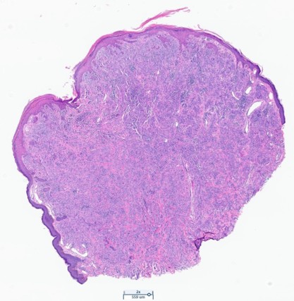
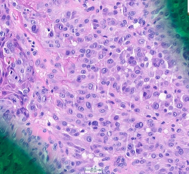
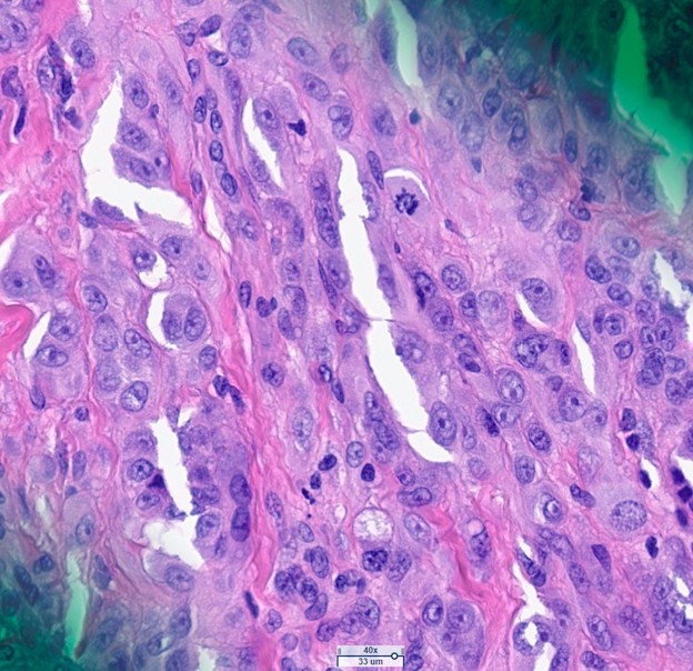
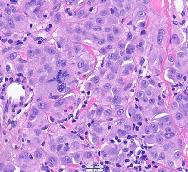
The biopsy showed a nodular, dermal, melanocytic proliferation (Figure 1) composed of epithelioid cells with moderate to severe cytologic atypia (Figure 2). The cells show prominent nucleoli, nuclear pleomorphism, nuclear grooves and other chromatin irregularities. Scattered atypical mitotic figures were observed (up to 2 per mm2, Figures 3-4). There was focal ulceration above the lesion and numerous lymphocytes scattered among the lesional cells in the dermis. The lesional cells extended to the margins of resection.
Immunohistochemistry
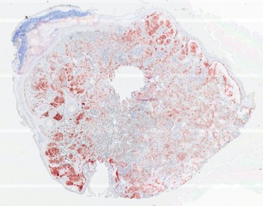
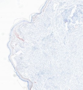
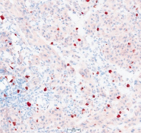
Lesional cells were positive for Melan-A, S100, and PRAME, but negative for HMB45. Lesional cells also showed loss of p16. Ki67 index was low (<5%).
Molecular Pathology and Cytogenetics
Further molecular testing was performed on the lesion. Fluorescent in situ hybridization (FISH) testing showed homozygous loss of CDKN2A in lesional cells. Testing for c-MYC, CCND1, and RREB1 did not show alterations.
Oncomine Comprehensive panel testing did not identify any pathogenic mutations, including TERT alterations.
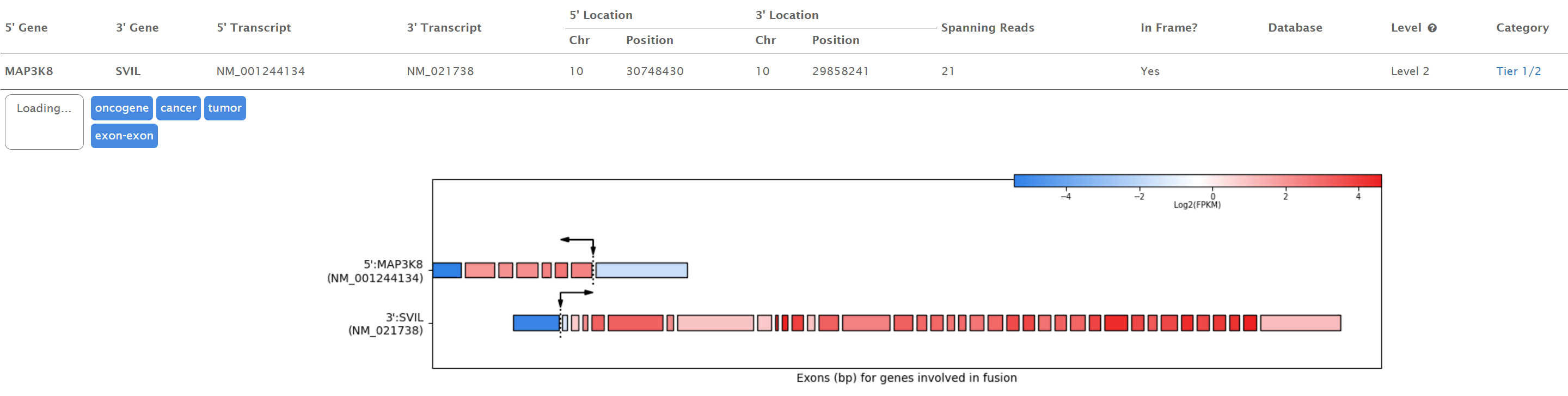
RNA-sequencing analysis of the tumor revealed a fusion between MAP3K8::SVIL.