
The Informatics Division at UPMC is one of the premier informatics groups in the United States with a national and international reputation. The division supports clinical practice, is involved in cutting edge research, training, and actively participates in commercial product development with UPMC Enterprises. Dr. Liron Pantanowitz is the Chair of the Department of Pathology and Maud L. Menten Professor of Pathology, Dr. Matthew Hanna is the Vice Chair for Pathology Informatics, Dr. Hooman Rashidi serves as the Executive Vice Chair of Computational Pathology and Dr. Ibrahim Abukhiran serves as the Director of the Pathology Image Analysis Laboratory. Additional informatics faculty include Drs. Sarah Wheeler, Thomas Pearce, Octavia Palmer, Jeffrey Fine, Kenichi Tamama, Suzanne Bakdash, and Mana Mohebnasab.
 The division is instrumental in providing integrated lab information system services and other key informatics services such as digital imaging and regulatory compliance to support patient care, education and training at UPMC. The division provides oversight for Sunquest for clinical laboratories, CoPath for anatomical pathology, Clarity LIMS and MGP client portal for molecular pathology, and several other specialty lab systems (e.g. Vitalent, HistoTrac). The division's faculty work closely with the Information Service Division (ISD) of UPMC to maintain, customize and develop commercial and niche clinical applications, instrument interfaces, and automation for the laboratory in various UPMC hospitals. The division is actively involved in providing informatics and analytics tools to help standardize pathology practice across all hospitals in the UPMC system and to enable evidence-based practice improvements. The division also converts lab information systems for newer labs joining the UPMC network. Teleconsultation service is a key priority with national and international business partners. We accept digital consults from around the world via our consultation portal https://pathconsult.upmc.com/ Faculty are engaged in developing cutting edge informatics tools related to whole slide imaging, image analysis algorithms, artificial intelligence and molecular informatics. The Pathology Informatics Division works closely with the Department of Biomedical Informatics at the University of Pittsburgh https://www.dbmi.pitt.edu The Informatics faculty also serve on key committees for several pathology organizations (API, DPA, CAP, USCAP, ASC, AMP, ADLM, ASCP) and journal editorial boards (such as JPI).
Division Clinical Activities
The informatics division maintains and develops cutting-edge clinical information systems to support anatomic pathology, clinical pathology, hematopathology, and molecular pathology. Over seventy percent of all the information generated in an academic or community hospital is derived from the activities of clinical and anatomic pathology. Our division manages this enormous flow of information and turns it into accessible knowledge for use by clinicians.
Anatomical Pathology
The division maintains and develops clinical information systems to support all types of pathology practice (e.g., subspecialty academic or general community). Since 2012 we have been using an internally developed web portal to perform international teleconsultations. Since that time, we have performed over 5000 international consultations. The Anatomic Pathology informatics team manages the AP-LIS which is a highly integrated lab information system deployed across multiple sites that maintains the anatomical pathology digital archive, handles coding, imaging, and electronic reporting. Our pathology reports are distributed to all of UPMC Electronic Health Records, enterprise data warehouse, and research digital warehouses. The Division customized barcoding for consults, automated several quality assurance tasks, and implemented SAFER codes.
Digital Pathology
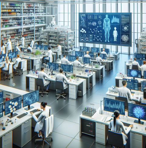 The informatics division has a fully deployed digital pathology system for scanning that is integrated with the AP-LIS. We scan thousands of slides every month and utilize WSIs for clinical work, education, and research. A dozen hybrid WSI-live robotic scanners have been deployed for intraoperative consultations via telepathology. UPMC's Pathology Image Analysis Laboratory, directed by Dr. Ibrahim Abukhiran, aims to enhance diagnostic accuracy and patient outcomes by advancing clinical image analysis through innovative techniques. Established in 2018 and accredited by the College of American Pathologists, our laboratory focuses on developing and validating image analysis algorithms, integrating these with clinical data, and collaborating with clinicians and researchers to apply findings in practice. Unique to UPMC, our laboratory features clinically validated algorithms for various cancers, including breast biomarker quantification, CD8 positive tumor infiltrating lymphocytes quantification for colon cancer, melanoma, head and neck squamous cell carcinoma, and metastatic carcinoma of any primary origin for immunotherapy selection. We employ a distinctive testing methodology with rigorous quality control measures. Supported by an extensive digital infrastructure, we offer services beyond UPMC, including slide digitization and quantitative image analysis.
The digital pathology core also partners with the Pitt Biospecimen Core to provide imaging services for researchers. http://www.pittbiospecimencore.pitt.edu/imaging-services.
Clinical Pathology
For Clinical Pathology, the division supervises the maintenance of the CP-LIS software that coordinates electronic data generated by a large number of lab instruments, manages a sophisticated total automation system, and feeds results of millions of lab tests into the EHR. The division successfully tackles novel informatics projects such as the creation of segregated VLAN infrastructure for enhanced LIS security and IT Big Bang across the UPMC network hospitals. In collaboration with the clinical analytics team, the pathology informatics division is currently working on Big Data laboratory analytics, and data democratization to enable evidence-based practice and workflow improvements.
Molecular Pathology
The Division of Molecular and Genomic Pathology (MGP) has pioneered in molecular informatics and using modern IT infrastructure for deployment of novel, large, and complex information systems such as Clarity LIMS and development of state-of-the-art enterprise apps, such as the MGP consultation portal.
Research Activities
The research mission of the division is to develop cutting-edge medical informatics, developmental and research programs focused on next generation pathology reporting methods, imagery, and genomics bioinformatics. Our division is extremely productive and has published many articles, textbook chapters and books. Areas of active research include international telepathology, image analysis, next generation sequencing informatics, high-throughput and cloud computing architectures, 3D imaging, in vivo/ex vivo microscopy, and artificial intelligence (AI) including explainable AI (xAI). The division collaborates with many physicians, scientists and industry to support their research initiatives. The Division has also started a Digital Pathology Research Center, and has collaborations with the Center of Computational Pathology and AI Center of Excellence.
Education and Training
The division directs an annual pathology informatics training course for pathology residents and oversees resident electives in pathology informatics at UPMC. We follow the Pathology Informatics Essentials for Residents (PIER) curriculum. The division also provides updated online resources (e.g. recorded lectures, wiki) for pathology trainees. Our fellowship in pathology informatics is highly successful with many high quality candidates applying.
Selected Publications by Division Faculty in Informatics
Articles
- Hanna M, Pantanowitz L, Jackson B, Palmer O, Visweswaran S, Pantanowitz J, Deebajah M, Rashidi H. Ethical and Bias Considerations in Artificial Intelligence (AI)/Machine Learning. Mod Pathol. 2024 Dec 16:100686. doi: 10.1016/j.modpat.2024.100686. Epub ahead of print. PMID: 39694331.
- Pantanowitz L, Pearce T, Abukhiran I, Hanna M, Wheeler S, Soong TR, Tafti AP, Pantanowitz J, Lu MY, Mahmood F, Gu Q, Rashidi HH. Non-Generative Artificial Intelligence (AI) in Medicine: Advancements and Applications in Supervised and Unsupervised Machine Learning. Mod Pathol. 2024 Dec 13:100680. doi: 10.1016/j.modpat.2024.100680. Epub ahead of print. PMID: 39675426.
- Pantanowitz J, Manko CD, Pantanowitz L, Rashidi HH. Synthetic Data and its Utility in Pathology and Laboratory Medicine. Lab Invest. 2024 Jun 24:102095. doi: 10.1016/j.labinv.2024.102095. Epub ahead of print. PMID: 38925488.
- Rashidi HH, Fennell BD, Albahra S, Hu B, Gorbett T. The ChatGPT conundrum: Human-generated scientific manuscripts misidentified as AI creations by AI text detection tool. J Pathol Inform. 2023 Oct 17;14:100342. doi: 10.1016/j.jpi.2023.100342. PMID: 38116171; PMCID: PMC10727991.
- Hanna MG, Olson NH, Zarella M, Dash RC, Herrmann MD, Furtado LV, Stram MN, Raciti PM, Hassell L, Mays A, Pantanowitz L, Sirintrapun JS, Krishnamurthy S, Parwani A, Lujan G, Evans A, Glassy EF, Bui MM, Singh R, Souers RJ, de Baca ME, Seheult JN. Recommendations for Performance Evaluation of Machine Learning in Pathology: A Concept Paper From the College of American Pathologists. Arch Pathol Lab Med. 2023 Dec 2.
- Fortman D, Karunamurthy A, Hartman D, Wang H, Seigh L, Abukhiran I, Najjar YG, Pantanowitz L, Zarour HM, Kirkwood JM, Davar D. Automated Quantitative CD8+ Tumor-Infiltrating Lymphocytes and Tumor Mutation Burden as Independent Biomarkers in Melanoma Patients Receiving Front-Line Anti-PD-1 Immunotherapy. Oncologist. 2024 Jul 5;29(7):619-628.
- Elkhadrawi M, Lopez-Nunez O, Akcakaya M, Wheeler SE. Improving Lyme disease testing with data driven test design in pediatrics. J Pathol Inform. 2023 Feb 16;14:100300.
- Vizcarra JC, Pearce TM, Dugger BN, Keiser MJ, Gearing M, Crary JF, Kiely EJ, Morris M, White B, Glass JD, Farrell K, Gutman DA. Toward a generalizable machine learning workflow for neurodegenerative disease staging with focus on neurofibrillary tangles. Acta Neuropathol Commun. 2023 Dec 18;11(1):202.
- Hanna MG, Ardon O, Reuter VE, Sirintrapun SJ, England C, Klimstra DS, Hameed MR. Integrating digital pathology into clinical practice. Mod Pathol. 2022 Feb;35(2):152-164. doi: 10.1038/s41379-021-00929-0. Epub 2021 Oct 1. Erratum in: Mod Pathol. 2022 Feb;35(2):287.
- Elkhadrawi M, Stevens BA, Wheeler BJ, Akcakaya M, Wheeler S. Machine Learning Classification of False-Positive Human Immunodeficiency Virus Screening Results. J Pathol Inform. 2021 Nov 20;12:46
- Landau MS, Pantanowitz L. Artificial intelligence in cytopathology: a review of the literature and overview of commercial landscape. J Am Soc Cytopathol. 2019; 8(4):230-241.
- Pearce TM, Nikiforova MN, Roy S. Interactive Browser-Based Genomics Data Visualization Tools for Translational and Clinical Laboratory Applications. J Mol Diagn. 2019 Nov;21(6):985-993.
- Pantanowitz L, Preffer F, Wilbur DC. Advanced imaging technology applications in cytology. Diagnostic Cytopathology. 2019; 47:5-14.
- Tizoosh HR, Pantanowitz L. Artificial intelligence and digital pathology: challenges and opportunities. Journal of Pathology Informatics. 2018; 9:38
- Fragetta F, Garcia-Rojo M, Baidoshvili A, Evans A, Yagi Y, Tuthill M, Hartman D, Fukya J, Pantanowitz L. The importance of eSlide macro images for primary diagnosis with whole slide imaging. J Pathol Inform. 20189; 9:46
- Pantanowitz L, Carter A, Kurc T, Sharma A, Sussman A, Saltz J. 20 Years of Digital Pathology: an overview of the road travelled, what is on the horizon, and emergence of vendor neutral archives. J Pathol Inform. 2018; 9:46.
- Evans AJ, Bauer TW, Bui MM, Cornish TC, Duncan H, Glassy EF, Hipp J, McGee RS, Murphy D, Myers C, O'Neill DG, Parwani AV, Rampy BA, Salama ME, Pantanowitz L. US Food and Drug Administration approval of whole slide imaging for primary diagnosis: a key milestone is reached and new questions are raised. Archives of Pathology & Laboratory Medicine. 2018; 142:1383-1387.
- Hartman DJ, Ferris R, Rimm D, Ahmed F, Pantanowitz L. Utility of CD8 score by automated quantitative image analysis in head and neck squamous cell carcinoma. Oral Oncology 2018; 86:278-287.
- Hanna M, Nine J, Ishtiaque A, Pantanowitz L. Augmented reality technology using Microsoft HoloLens in anatomic pathology. Archives of Pathology & Laboratory Medicine. 2018; 142:638-644.
- Niazi MK, Hartman DJ, Pantanowitz L, Gurcan M. Identifying tumor in pancreatic neuroendocrine neoplasms from Ki67 images using transfer learning. PLOS ONE. 2018; 13(4):e0195621.
- Pearce TM, Nikiforova MN, Roy S. Interactive Web-Based Genomics Data Visualization Tools for Translational and Clinical Laboratory Applications. J Mol Diagn. 2019 Aug 2. pii: S1525-1578(19)30348-4. doi: 10.1016/j.jmoldx.2019.06.005. [Epub ahead of print]
- Callenberg KM, Santana-Santos L, Chen L, Ernst WL, Barbi De Moura M, Nikiforov YE, Nikiforova MN, Roy S. Clinical Implementation and Validation of Automated HGVS Nomenclature System for NGS-based Assays for Cancer. J Mol Diagn. 2018 Sep;20(5):628-634.
- Roy S, Coldren C, Karunamurthy A, Kip NS, Klee EW, Lincoln SE, Leon A, Pullambhatla M, Temple-Smolkin RL, Voelkerding KV, Wang C, Carter AB. Standards and Guidelines for Validating Next-Generation Sequencing Bioinformatics Pipelines: A Joint Recommendation of the Association for Molecular Pathology and College of American Pathologists. J Mol Diagn. 2018 Jan;20(1):4-27.
- Roy S, LaFramboise WA, Liu T, Cao D, Luvison A, Miller C, O'Sullivan RJ, Zureikat AH, Melissa E. Hogg ME, Tsung A, Lee KK, Bahary N, Brand RE, McGrath K, Nikiforova MN, Slivka A, Zeh HJ, Singhi AD. The Metastatic Progression of Pancreatic Neuroendocrine Tumors is Characterized by Recurrent Genetic Alterations in Chromatin Remodeling Genes and CDKN2A. Gastroenterology. 2018 Feb 24. pii: S0016-5085(18)30243-9. doi: 10.1053/j.gastro.2018.02.026. [Epub ahead of print].
- Nguyen L, Tosun AB, Fine J, Lee A, Taylor DL, Chennubhotla C. Spatial statistics for segmenting histological structures in H&E stained tissue images. IEEE Trans Med Imaging. 2017 Mar 16. doi: 10.1109/TMI.2017.2681519.
- Farahani N, Liu Z, Jutt D, Fine JL. Pathologists' Computer-Assisted Diagnosis: A Mock-up of a Prototype Information System to Facilitate Automation of Pathology Sign-out. Arch Pathol Lab Med. 2017;141:1413-1420. doi: 10.5858/arpa.2016-0214-OA).
- Nguyen L, Tosun AB, Fine JL, Taylor DL, Chennubhotla SC. ARCHITECTURAL PATTERNS FOR DIFFERENTIAL DIAGNOSIS OF PROLIFERATIVE BREAST LESIONS FROM HISTOPATHOLOGICAL IMAGES. 2017 IEEE International Symposium on Biomedical Imaging (ISBI 2017).
- Spagnolo DM, Gyanchandani R, Al-Kofahi Y, Stern AM, Lezon TR, Gough A, Meyer DE, Ginty F, Sarachan B, Fine J, Lee AV, Taylor DL, Chennubhotla SC. Pointwise mutual information quantifies intratumor heterogeneity in tissue sections labeled with multiple fluorescent biomarkers. J Pathol Inform. 2016;7:47.
- Roy S, LaFramboise WA, Nikiforov YE, Nikiforova MN, Routbort MJ, Pfeifer J, Nagarajan R, Carter AB, Pantanowitz L. Next Generation Sequencing Informatics: Challenges and Strategies for Implementation in a Clinical Environment. Arch Pathol Lab Med. 2016 Sep;140(9):958-75.
- Roy S, Pfeifer JD, LaFramboise WA, Pantanowitz L. Molecular digital pathology: progress and potential of exchanging molecular data. Expert Rev Mol Diagn. 2016 Sep;16(9):941-7.
Books
- Pantanowitz L, Balis UJ, Tuthill JM. Pathology Informatics: Theory & Practice. ASCP Press. 2012.
- Pantanowitz L, Parwani AV. Digital Pathology. ASCP Press. 2017.
- de Baca ME, Spinosa JC, Aller R, Badizadegan K, Blouin AG, Castellani W, Chen P, Gilbertson J, Harrison J, Henricks W, Kennedy M, Knapik C, Pantanowitz L, Reichard RR, Robb J, Stram M. CAP Pathology resource guide: Clinical Informatics. Version 1.2.0.0. Northfield, IL: College of American Pathologists; 2018.
- Hipp J, Bauer TW, Bui MM, Cornish TC, Evans AJ, Glassy EF, Murphy D, Pantanowitz L, Parwani AV, Rampy BA, Salama ME, Water R. CAP Pathology Resource Guide: Digital Pathology. Version 6.0(1). Northfield, IL. College of American Pathologists. 2016.
- Fitzmaurice M, Fine JL, Fischer AH, Kaufman JH, Mathur SC, Mirkovic J, Rao BK, Shevchuk MM, Sussman LK, Vajpeyi R, editors. In vivo microscopy resource guide. Version 7.0. Northfield, IL: College of American Pathologists; 2016.
- Pantanowitz L, Parwani AV. Practical Informatics for Cytopathology. Springer. 2014.